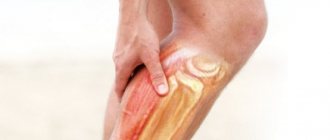
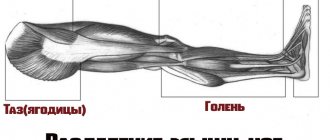
Candice Swanepoel Katya Egorova, coach of Reboot sports studios:
“Before you start stretching your calf muscles, remember a few simple rules:
- it must be performed when you are already well warmed up, ideally at the end of the lesson;
- no jerks or sudden movements, do everything smoothly;
- Pay close attention to your internal sensations: if you feel unbearable pain, then reduce the amplitude of the exercise or stop doing it altogether.
Another nuance that should not be ignored is cramps or spasms in the calf muscles. Most often, this is a signal from the body about a lack of magnesium. Don’t be lazy to take tests to make sure of this and choose the vitamin complex you need. Plus, add magnesium-rich foods to your diet, such as beans, lentils, bananas, avocados, figs and nuts. In addition, try to drink at least 1.5 liters of water per day: dehydration can also lead to cramps.
Candice Swanepoel I also remind you that the condition of the calf muscles is adversely affected by excessive loads on the legs, flat feet and uncomfortable shoes. Therefore, give your feet a rest (in the evening before going to bed, lie down for a few minutes with your legs raised up), do not sit cross-legged (this interferes with blood circulation), wear comfortable shoes or carry a change of clothes with you and replace regular slippers with orthopedic ones.”
Examination of the child at the reception
At a medical consultation, a complete examination of the child is required, with an assessment of the following factors while standing, sitting and lying down:
- Constitutional features of development,
- The presence of markers of possible undifferentiated (most often) connective tissue dysplasia,
- The manifestation of hereditary factors influencing the individual development of the foot, lower extremities,
- Functional anatomical features of the installation of the child’s foot in the phases of supporting and unsupported walking,
- Plantoscopy results,
- Individual characteristics of shoe wear,
- Analysis of submitted medical documents and examinations,
- If there are siblings, a comparative analysis of all the above factors in their family totality is carried out.
To prescribe exercises that develop the tone-strength characteristics of the arch-forming muscles of a child’s foot, a preliminary medical consultation is advisable.
| Rice. 1 | Figure 1 shows in an artificially hypertrophied form two different installations of the foot and knee joint - valgus (a) and varus (b). The reasons for such features are considered by the doctor together with the parents, with a mandatory analysis of all possible causative factors. It is important to emphasize that both forms of installation of the feet and knee joints can be a consequence of the child getting into bed too early. The same picture can be observed as a temporary phenomenon in premature babies, because the bone tissue has not “accumulated” enough calcium hydroxyapatite salts with a sufficient amount of collagen - the bone matrix that gives children’s bones some flexibility and elasticity. |
| Rice. 2 | Figure 2 shows the valgus position of the foot (a) on the supporting surface with the peak points of gravitational loads shifted to the inner part of the forefoot and heel (b) which have their own specific negative consequences for the growing foot and the musculoskeletal system of the child. |
| Rice. 3 | Figure 3 shows the extreme degree of combined longitudinal and transverse flatfoot with complete loss of all arches of the foot and expressed by diffuse gravitational overload of the entire inner part of the foot. Such a foot lacks the ability to absorb shock and adapt to motor loads. The load on the overlying parts of the musculoskeletal system changes, the gait becomes flopping, and pain often occurs in the area of the ligamentous-tendon system of the foot and ankle; The knee joint, connected to the foot in a single functional chain, also suffers. |
| Fig 3.1 | In Fig. Figure 3.1 shows the deformation (flattening) of the arch of the transverse arch of the foot, characteristic of older children and adolescents; The points of peak gravitational loads are shifted and concentrated in the center of the forefoot. This is the site of callus development; gait changes, especially in the phase of supporting walking with the load of the anterior section and separation from the support. Pain syndrome is also formed in this place. |
| Rice. 4 | Rice. 5 | Rice. 6 |
To better understand the features of the functional anatomy of the foot, it is worth considering examples of the so-called tendon stapes of the foot, formed by the intersection of the tendons of the long peroneus muscle and the tibialis posterior muscle - the main statodynamic support of the longitudinal arches of the foot (Fig. 4, 5, 6). It is these muscles that are very weak in children and adolescents with flat feet, and it is these muscles that need to be trained to achieve proper support for the skeletal structure of the foot.
Rice. 7
Figure 7 shows a side view of the foot. So-called passive tightening of the arches of the feet are presented. These are elements of the ligamentous apparatus of the foot, which forms and maintains its spatial configuration and is most often determined by hereditary factors.
Foot exercises
Figure 8-16 shows simple and accessible exercises for independent development of your own foot muscles and the muscles that come to the foot with their tendons from the lower leg.
| Rice. 8 |
| Rice. 10 |
| Rice. eleven |
|
|
|
|
|
Workout 2: Challenge
I recommend training your calves twice a week. But don't consider this training as something of secondary importance. Treat your calves the same way you treat your biceps, triceps, or chest muscles. In other words, train them constantly and intensely.
Workout 2 is short but painful. It consists of two giant approaches. In many gyms, calf training machines are located next to each other. You will need three different machines. You can use a donkey calf raise machine, a seated calf raise machine, a standing calf raise machine, a leg press machine (to perform calf raises), and/or an angled calf raise machine. If your gym only has two of the machines listed above, you can perform the single-leg raises described above as a third exercise.
According to this program, you perform the first exercise, then immediately jump to another machine and perform the next exercise, after completing which you immediately move on to the third exercise. For each set, choose a weight with which you can perform at least 10 repetitions, but always perform the exercise to muscle failure. It is necessary to prepare these three machines in advance before performing this superset so that you do not have to stop to adjust the weight. When you finish the first superset, your calves will hurt like crazy. This is what you need.
Jason, my calves are killing me!
Without a doubt, if you train your calves correctly, with the required intensity, they will grow. You'll curse me while you're in the shower and on the car ride home, but it's worth it. The reward for having tears streaming down your face after completing a set of 50 reps is simple. Firstly, you will have increased psychological strength to overcome the pain barrier. Secondly, your calves will be the envy and admiration of everyone in the gym. I don't know a more impressive sight in the gym than a pair of huge calves. Those who train their calves intensively cannot be confused with those who do it casually.
Upper back stretch
Kneel on the floor and lower your buttocks until you touch your heels. Extend your arms forward and touch the floor with your palms and forehead. Stretch forward, feel the stretch in the thoracic spine.
Why is it important to do this exercise: long sitting work and long walking lead to tension in the thoracic spine. Stretching the upper back helps relieve stress in this area.
Clinical picture
Patients with heel spurs complain of pain when taking their first steps in the morning, after sitting at a desk or driving for a long time - the so-called “starting pain”. The pain is felt in the heel and can be quite severe (Fig. 1).
Often improvement occurs after the first steps or stretching the muscles of the lower leg and fascia of the foot. However, the pain usually returns during the day, especially if the patient walks or stands a lot. Burning pain is not typical for plantar fasciitis and may occur due to nerve irritation (eg, Baxter's neuritis).
The main reasons for the development of plantar fasciitis:
- age
- recent increase in physical activity level (eg, a new running program)
- work that requires standing for long periods of time
- weight gain
- tight (rigid) calf muscles
On clinical examination, pain is most often localized along the inner surface of the heel from the plantar surface of the foot. Pain also occurs with direct pressure (palpation) on the specified area.
Stiffness in the lower leg muscles is also a common symptom. Symptoms may be aggravated by pulling the toes toward you, thereby stretching the plantar fascia (see Figure 3). There is a connection between flat feet and the development of plantar fasciitis, but this disease can develop in any type of foot.
Plantar fasciitis is the most common cause of heel pain, but there are other less common causes:
- pain associated with heel overload
- atrophy of the soft tissues of the foot
- entrapment of the first branch of the lateral plantar nerve (Baxter's nerve)
- tarsal tunnel syndrome
- calcaneal stress fracture
- inflammation of the periosteum
- inflammation caused by seronegative arthritis
Additional research methods
The diagnosis of plantar fasciitis is usually made based on patient complaints and clinical examination. X-rays of the feet are not necessary to make a diagnosis. However, if an x-ray is nevertheless prescribed, a heel spur is visualized on the lateral projections.
It is important to understand that overload of the plantar fascia can cause excessive bone formation, in the form of heel spurs. However, the presence of a heel spur does not directly correlate with symptoms.
In many patients, a spur is visible on radiographs of the foot, but there are no symptoms, and vice versa - in patients suffering from plantar fasciitis, a heel spur is absent on radiographs.
Initially, patients are not prescribed MRI of the foot. However, if symptoms do not improve after treatment, an MRI may be ordered to rule out other causes of heel pain, such as a stress fracture of the calcaneus.
Front thigh stretch
Stand on your right leg, bend your left knee and take it back. Hold your left ankle with your left hand, slightly pulling it up. You should feel a pulling effect in your left thigh. Repeat for the other leg.
Why this exercise is important: Stretching the front of your thighs reduces muscle tension and prevents you from feeling stiff in the morning. The exercise also helps in preventing knee injuries.
Tips for training calves
After reading this, you will definitely see people in the gym making the same mistakes that you are making now. Firstly, it depends on the shoes. Train your calf muscles in flat shoes. Even better is to train barefoot.
Secondly, foot placement is very important. Probably the biggest mistake people make when training calves is bending their knees too much. Remember that your goal is to exclude the quadriceps from the movement and isolate the calf muscles. It's okay to bend your legs slightly, but I think your legs should be as straight as possible. Place your feet so that the distance between your heels is wider than the distance between your toes. It is very important that the main load goes directly to the thumbs. Using weights will actually help achieve full contraction on each rep.
Another factor that is often overlooked is stretching. Before training my calves, I like to give them a good stretch. By the way, I have seen two different devices specifically designed to stretch the calf muscles. The calves consist of the soleus muscle and the gastrocnemius muscle. To stretch the soleus muscle, sit on a seated calf raise machine and stretch without using weights. To stretch the calf muscle, perform calf raises while standing, lowering your heels toward the floor. It's good if you stretch your calves often, between sets.
Ultimately, you should concentrate on achieving a full contraction on each rep. As you lower your heels, you will feel yourself sliding off the machine. At the top of the exercise, you will be standing almost on your thumbs alone. You may have already noticed that those who use huge weights and bounce with every rep have small calves.
Help and advice from experts
K. Ilkevich, fitness expert, personal bodybuilding trainer
The calf muscles are the most difficult to develop for 2 reasons: improper selection of training exercises and a passive lifestyle.
The best option for working your calves would be 1-2 sessions per week before your main workout. Use an average of 3 calf ligaments in 3 sets. He often advises combining different execution speeds until muscle failure. This workout will allow you to achieve maximum muscle growth without feeling overtrained.
V. Berlichev, professional bodybuilder, personal trainer
A large number of athletes pay very little attention to the development of the calf muscles. Therefore, quite often, even among athletes, we can observe disproportionate figures: the torso and arms look quite powerful, but the legs seem too thin and thin. The biggest mistake is the habit of leaving pumping your calves until the end of the workout - at this time the body will get tired, and you simply will not be able to work out your shins efficiently and slowly. For this reason, I always recommend loading your calves early in your workout.
L. Ulyanicheva, sports expert, professional fitness trainer
Girls should not be afraid to pay attention to the soleus muscles. With regular exercise, your legs will not become pumped up, but will look toned. One of the simplest exercises can be called seated leg raises. Use additional weight, doing at least 15 repetitions. Calves recover quite quickly, so reasonable weights will benefit them.
