The older a person gets, the more difficult it is to move due to disruption of large joints. This is due to the destruction of bone tissue and aging of the cartilage surface. When the hip joint is affected, these problems sharply limit a person’s capabilities, which dictates the need to search for relevant therapeutic measures.
Most often, it is precisely this type of joint destruction that patients begin treatment for. But it’s too late not only to do gymnastics, but also to do any other procedures.
Not everyone is ready to immediately realize that the only radical option for getting rid of pain and difficulty moving in the joint is hip replacement. One of the ways to restore the functionality of a joint with coxarthrosis is therapeutic exercises. Despite the fact that exercises cannot save the joint, they can help develop the ligamentous-muscular system, which will have a positive effect on the postoperative period.
The essence of physical training
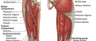
The main problem that activates the process of destruction of the hip joint is a decrease in the amount of synovial fluid. The glands of the shell of the same name, lining the articulation cavity, are responsible for its production. As soon as there is less synovial fluid, the mobility of the articular surfaces is impaired and the friction between them increases. This leads to a narrowing of the joint space, which in severe situations completely immobilizes the patient. This is how arthrosis of the hip joint occurs.
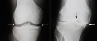
This is a real image of the femoral head with stage 2-3 arthrosis. Look carefully and think, will gymnastics help in this case?
Medicines can slow down the progression of the disease, but the process cannot be stopped. At an unfavorable moment, arthrosis will lead to serious pain, which will significantly affect the quality of life. However, even at the moment when it becomes clear that endoprosthetics surgery is no longer possible, therapeutic exercises will prove extremely useful for improving the functioning of the joint, both before and after surgery.
The effect of exercises carried out at home is as follows:
- stimulation of blood flow around the damaged joint;
- restoration of muscle performance and prevention of muscle atrophy;
- stimulation of the glands of the synovial membrane due to intensive blood supply;
- reducing the progression of arthrosis;
- stretching and mobilization of the ligamentous apparatus;
- general strengthening of the body and immune system.
After any gymnastic exercises, the mood improves, which leads to an increase in a person’s ability to fight the disease.
A network of blood vessels that supplies tissues with useful substances that promote rapid regeneration of the cartilage surface.
However, not only the psychological factor influencing the patient plays a role in activating restoration processes in the hip joint. There are also quite tangible changes that the patient will feel after treatment through regular exercise. These include:
- increased range of motion in the joint;
- pain relief;
- increased strength in a limb;
- easier walking;
- reduction in the frequency of colds;
- improving quality of life.
Although gymnastics will not radically change the situation, the joint will still have to be replaced with prosthetics; it will help reduce rehabilitation time and speedily restore motor activity in the postoperative period. This will be possible thanks to developed muscles and well-developed tendons, as well as stable blood flow in the hip joint.
There are many options for gymnastic exercises done at home. But the effectiveness of their treatment is completely different. The most relevant training is that proposed by a professional exercise therapy instructor, as well as the classic methods of Evdokimenko and Bubnovsky. A video of classes with a physical therapy specialist clearly shows the optimal set of exercises.
Minimally invasive endoprosthetics in the Czech Republic: doctors, rehabilitation, terms and prices.
Find out more
Who needs to train the pelvic floor muscles and why?
Even if you do not experience problems in the above areas, everyone needs to strengthen their muscle corset, since the vast majority suffer from a lack of physical activity and physical exercise. Taking care of muscle tone is especially necessary for women after the birth of a child who suffer from hemorrhoids, urinary incontinence, pain in the sacrum or lower back. It is necessary to exercise and strengthen the muscles, keep them in good shape so that the quality of life does not deteriorate. Strengthening the pelvic floor muscles is indispensable; if they are toned, it improves the quality of sexual life and prevents urinary incontinence or bladder prolapse. Women planning a pregnancy can perform a set of therapeutic exercises to tone their muscles, which will make childbirth less painful, and after it the young mother will quickly return to her previous shape.
Gymnastics Evdokimenko
Since the early 2000s, Dr. Evdokimenko’s practice of therapeutic exercises has become widespread in medical practice. Today he is a well-known rheumatologist, a specialist in joint restoration through physical training. Has the status of academician at one of the Moscow technical universities. His medical career began in 1984, when the future specialist received a paramedic education. The further period is associated with practical work and study at the institute, which Pavel Evdokimenko graduated from in 1994. From that moment on, he chose the direction of physical therapy for various osteo-articular pathologies as his specialization.
Pavel Evdokimenko.
Further work at the arthrosis center in Moscow, and then subsequent improvements at the Research Institute of Rheumatology of the Russian Academy of Medical Sciences, prompted the doctor to develop a set of exercises that help prolong the active function of the joints. In 2003, the first book was published, telling in detail about the method of Pavel Evdokimenko. Today, exercise is recognized throughout the world and helps slow the progression of coxarthrosis. Even in the most difficult situations, when surgical intervention is already inevitable, the use of therapeutic exercises can dramatically reduce postoperative rehabilitation.
Although gymnastics can heal arthrosis, it cannot completely replace the installation of a prosthesis. However, there are significant positive qualities that are discussed below.
- Strengthening the thigh muscles. The less their atrophy, the easier it is to move the limb. A strengthened muscle frame will help reduce the load on the painful joint, thereby slowing down the progression of arthrosis.
- Activation of blood flow in the synovium. Improving the blood supply to the affected hip helps reduce the rate of reduction of the joint space. This is achieved by increasing the production of synovial fluid. As a result, friction in the joint is reduced and functionality is increased.
- Reducing osteoporosis. Achieved by increasing active movements in the joint. The processes of rarefaction of bone tissue are reduced, as the work of osteoblasts is activated.
- Strengthening the back frame. Since physical training stimulates muscle activity not only in the thigh, but also in the buttock and lumbar region, the load on the affected joint and spine is further reduced. This makes movement easier and also relieves pain.
- Increasing joint resistance to physical stress. This is achieved through long training.
- Easy adaptation in the postoperative period during joint replacement. Since the muscles, as well as the ligamentous apparatus, are in an optimally developed state, even the first steps after surgery are easier to bear, and rehabilitation will be short and painless.
Gymnastics video:
All exercises according to Evdokimenko’s method are performed many times. No major changes in the joint will occur within a month of training, so you need at least six months. But the systematic use of gymnastics promotes significant clinical improvement. The patient will feel a significant improvement in walking, as well as a decrease in pain.
Basic recommendations and precautions
The need for moderate physical activity during pregnancy can be characterized by the motto “It is better to sit than to lie down. Better standing than sitting. It’s better to walk rather than stand.” Everything needs moderation. Organized physical activity in the absence of contraindications will not harm anyone, including pregnant women. If you don't want to exercise, you can swim. If you don't like swimming, there is breathing practice and yoga. When none of the above sparks enthusiasm, try dancing or walking more outdoors. You can choose a type of activity that will be useful and enjoyable at the same time.
The main recommendations are as follows:
- do not overheat during training - this has a negative effect on the child;
- drink more water to stimulate the removal of toxins and normalize metabolism;
- the optimal time for training is 2 hours after breakfast;
- avoid excessive stress. The presence of shortness of breath in the mother means that the baby receives less oxygen;
- control your well-being. The presence of pain in the lower abdomen is a signal to stop exercising;
- there is no point in overworking: 15 minutes is enough to stretch your muscles;
- try not to do exercises during periods when “critical days” were supposed to occur;
- do not forget about proper breathing and relaxation.
Pregnancy is a crucial period when a woman should take care of her body. If there is no evidence, there is no point in lying on the couch for 9 months. Physical education will help prepare the body for childbirth, reduce pain, improve metabolism and avoid gaining excess body weight.
List of exercises according to Evdokimenko
The author offers 10 of the most relevant exercises that will help quickly stabilize the joint. In total, the therapeutic gymnastics course includes more than 50 different exercise options, but Pavel Evdokimenko believes that it is impossible to do without the dozens of the most significant. The entire list of exercises for the treatment of coxarthrosis is presented below.
- Leg lift. A simple exercise that is performed at a slow pace. You need to start with the affected joint. Standing on a hard surface, you should raise your leg bent at the knee. Then hold it for 45 seconds, then slowly lower it. Repeat with the healthy leg. The frequency of execution is 2 times a day.
- Dynamic leg raises. Repeat the first exercise at a fast pace. You need to hold your leg suspended for no more than 2 seconds. Number of repetitions – 15. Multiplicity – twice a day.
- Lying leg raises. Starting position – with your back on the floor, legs slightly apart. You should slowly raise the limb bent at the knee and hold it for 60 seconds. One-time execution is enough. Then repeat at a dynamic pace, at least 15 times with each leg.
- Leg raises and extensions. The exercise is suitable only for trained people. While lying on your back, you need to raise your closed limbs, without bending, up, hold in this position for 5 seconds, then spread them apart without lowering them. Close your legs and place them on the floor. Repeat at least 7 times.
- Lateral leg raises. Starting position – lying on the right half of the body. The legs are one on top of the other. You should raise your left leg, hold it for 10 seconds, then lower it. Repeat 10 times, then roll over to your left side and do the exercise with your right leg.
- Lateral foot lift with rotation. Starting position: lying on your right side. The straightened left leg must be raised up as high as possible, and then rotate the foot clockwise. Lower the limb, repeat 10 times. Then turn over onto your left side and do the exercise with your right leg.
- Lifting the pelvis. Lying on your back, with your knees bent, you should rest your elbows on the floor and lift your buttocks above the surface with the strength of your limbs. Hold in this position for 20 seconds, then lower. Repeat 10 times. If you have physical capabilities, the speed of the exercise can be increased, as well as the number of approaches.
- Long lean forward. Starting position – sitting on the floor, legs straightened as much as possible and spread no more than 10 cm wide. The essence of the exercise is to reach your feet with your hands, pull your body forward and remain in this position for 1 minute. Daily exercises stretch the ligamentous apparatus. The frequency of execution is only 1 time per day.
- Seated leg raises. Starting position – sitting on a chair. The legs are lowered down and bent at the knee joints. It is necessary to raise the limbs one by one to the maximum possible height, but holding for no more than 5 seconds. The repetition rate is at least 15 times with each leg.
- Knee bends. This is a slightly difficult exercise to understand, but it’s easier to do it step by step. To begin, take a sitting starting position with your back against the wall. Legs are straightened as much as possible and spread shoulder-width apart. The next step is to bend the sore leg at the knee joint, but do not lift the heel off the floor. Then reach the knee joint with your hands and smoothly, rocking it a little, try to bring it as close as possible to the healthy leg. There will be discomfort in the hip joint, but then you should relax and contract the thigh muscles. The pain will go away quickly. Repeat up to 10 times once a day.
All exercises are done daily, so it is important to set aside time and not miss classes. Within a month of training, tangible results will appear. If you need to undergo hip replacement surgery, then this duration of the course of exercises is sufficient for excellent preparation for surgical manipulation. If, on the advice of a doctor, endoprosthetics can be postponed, then classes should be continued for up to six months. The ideal option is one in which daily training is done for life. A video of all of Pavel Evdokimenko’s exercises will give a clear idea of the technique for performing them.
Minimally invasive endoprosthetics in the Czech Republic: doctors, rehabilitation, terms and prices.
Find out more
Conclusion
Hip dysplasia in children is a common pathology, but in the vast majority of cases it resolves on its own and does not require special medical intervention. In more complex cases, it may be necessary to wear various fixation devices for several weeks - until normal formation of the joint is completed - without requiring either casting or "strict" swaddling. In isolated cases, surgery may be required, and the earlier it is performed, the higher the chances of success. References
1. Wedge JH, Wasylenko MJ. The natural history of congenital dislocation of the hip: a critical review // Clin Orthop Relat Res 1978; :154. 2. Wedge JH, Wasylenko MJ. The natural history of congenital disease of the hip // J Bone Joint Surg Br 1979; 61-B:334. 3. Crawford AH, Mehlman CT, Slovek RW. The fate of untreated developmental dislocation of the hip: long-term follow-up of eleven patients // J Pediatr Orthop 1999; 19:641. 4. Bialik V, Bialik GM, Blazer S, et al. Developmental dysplasia of the hip: a new approach to incidence // Pediatrics 1999; 103:93. 5. Castelein RM, Sauter AJ. Ultrasound screening for congenital dysplasia of the hip in newborns: its value // J Pediatr Orthop 1988; 8:666. 6. Terjesen T, Holen KJ, Tegnander A. Hip abnormalities detected by ultrasound in clinically normal newborn infants // J Bone Joint Surg Br 1996; 78:636. 7. Marks DS, Clegg J, al-Chalabi AN. Routine ultrasound screening for neonatal hip instability. Can it abolish late-presenting congenital dislocation of the hip? // J Bone Joint Surg Br 1994; 76:534. 8. Dezateux C, Rosendahl K. Developmental dysplasia of the hip // Lancet 2007; 369:1541. 9. Harris NH, Lloyd-Roberts GC, Gallien R. Acetabular development in congenital dislocation of the hip. With special reference to the indications for acetabuloplasty and pelvic or femoral realignment osteotomy // J Bone Joint Surg Br 1975; 57:46. 10. Schwend RM, Pratt WB, Fultz J. Untreated acetabular dysplasia of the hip in the Navajo. A 34 year case series followup // Clin Orthop Relat Res 1999; :108. 11. Wood MK, Conboy V, Benson MK. Does early treatment by abduction splintage improve the development of dysplastic but stable neonatal hips? // J Pediatr Orthop 2000; 20:302. 12. Shaw BA, Segal LS, SECTION ON ORTHOPAEDICS. Evaluation and Referral for Developmental Dysplasia of the Hip in Infants // Pediatrics 2016; 138. 13. Murphy SB, Ganz R, Müller ME. The prognosis in untreated dysplasia of the hip. A study of radiographic factors that predict the outcome // J Bone Joint Surg Am 1995; 77:985. 14. Terjesen T. Residual hip dysplasia as a risk factor for osteoarthritis in 45 years follow-up of late-detected hip dislocation // J Child Orthop 2011; 5:425. 15. Lindstrom JR, Ponseti IV, Wenger DR. Acetabulular development after reduction in congenital dislocation of the hip // J Bone Joint Surg Am 1979; 61:112. 16. Cherney DL, Westin GW. Acetabular development in the infant's dislocated hips // Clin Orthop Relat Res 1989; :98. 17. Brougham DI, Broughton NS, Cole WG, Menelaus MB. The predictability of acetabular development after closed reduction for congenital dislocation of the hip // J Bone Joint Surg Br 1988; 70:733. 18. Albinana J, Dolan LA, Spratt KF, et al. Acetabular dysplasia after treatment for developmental dysplasia of the hip. Implications for secondary procedures // J Bone Joint Surg Br 2004; 86:876. 19. Weinstein SL, Mubarak SJ, Wenger DR. Developmental hip dysplasia and dislocation: Part II // Instr Course Lect 2004; 53:531. 20. US Preventive Services Task Force. Screening for developmental dysplasia of the hip: recommendation statement // Pediatrics 2006; 117:898. 21. Lorente Moltó FJ, Gregori AM, Casas LM, Perales VM. Three-year prospective study of developmental dysplasia of the hip at birth: should all dislocated or dislocatable hips be treated? // J Pediatr Orthop 2002; 22:613. 22. Larson JE, Patel AR, Weatherford B, Janicki JA. Timing of Pavlik harness initiation: Can we wait? // J Pediatr Orthop 2022. 23. Flores E, Kim HK, Beckwith T, et al. Pavlik harness treatment may not be necessary for all newborns with ultrasonic hip dysplasia // J Pediatr Health Care 2016; 30:304. 24. Rosendahl K, Dezateux C, Fosse KR, et al. Immediate treatment versus sonographic surveillance for mild hip dysplasia in newborns // Pediatrics 2010; 125:e9. 25. Burger BJ, Burger JD, Bos CF, et al. Frejka pillow and Becker device for congenital dislocation of the hip. Prospective 6-year study of 104 late-diagnosed cases // Acta Orthop Scand 1993; 64:305. 26. Atar D, Lehman WB, Tenenbaum Y, Grant AD. Pavlik harness versus Frejka splint in treatment of developmental dysplasia of the hip: bicenter study // J Pediatr Orthop 1993; 13:311. 27. Danielsson L, Hansson G, Landin L. Good results after treatment with the Frejka pillow for hip dysplasia in newborn infants: a 3-year to 6-year follow-up study // J Pediatr Orthop B 2005; 14:228. 28. Czubak J, Piontek T, Niciejewski K, et al. Retrospective analysis of the non-surgical treatment of developmental dysplasia of the hip using Pavlik harness and Frejka pillow: comparison of both methods // Ortop Traumatol Rehabil 2004; 6:9. 29. Carmichael KD, Longo A, Yngve D, et al. The use of ultrasound to determine timing of Pavlik harness discontinuation in treatment of developmental dysplasia of the hip // Orthopedics 2008; 31. 30. Tiruveedhula M, Reading IC, Clarke NM. Failed Pavlik harness treatment for DDH as a risk factor for avascular necrosis // J Pediatr Orthop 2015; 35:140. 31. Hedequist D, Kasser J, Emans J. Use of an abduction brace for developmental dysplasia of the hip after failure of Pavlik harness use // J Pediatr Orthop 2003; 23:175. 32. Cashman JP, Round J, Taylor G, Clarke NM. The natural history of developmental dysplasia of the hip after early supervised treatment in the Pavlik harness. A prospective, longitudinal follow-up // J Bone Joint Surg Br 2002; 84:418. 33. Nakamura J, Kamegaya M, Saisu T, et al. Treatment for developmental dysplasia of the hip using the Pavlik harness: long-term results // J Bone Joint Surg Br 2007; 89:230. 34. Walton MJ, Isaacson Z, McMillan D, et al. The success of management with the Pavlik harness for developmental dysplasia of the hip using a United Kingdom screening program and ultrasound-guided supervision // J Bone Joint Surg Br 2010; 92:1013. 35. Lerman JA, Emans JB, Millis MB, et al. Early failure of Pavlik harness treatment for developmental hip dysplasia: clinical and ultrasound predictors // J Pediatr Orthop 2001; 21:348. 36. Kitoh H, Kawasumi M, Ishiguro N. Predictive factors for unsuccessful treatment of developmental dysplasia of the hip by the Pavlik harness // J Pediatr Orthop 2009; 29:552. 37. Ömeroğlu H, Köse N, Akceylan A. Success of Pavlik Harness Treatment Decreases in Patients ≥ 4 Months and in Ultrasonographically Dislocated Hips in Developmental Dysplasia of the Hip // Clin Orthop Relat Res 2016; 474:1146. 38. Eidelman M, Katzman A, Freiman S, et al. Treatment of true developmental dysplasia of the hip using Pavlik's method // J Pediatr Orthop B 2003; 12:253. 39. Bialik GM, Eidelman M, Katzman A, Peled E. Treatment duration of developmental dysplasia of the hip: age and sonography // J Pediatr Orthop B 2009; 18:308. 40. Murnaghan ML, Browne RH, Sucato DJ, Birch J. Femoral nerve palsy in Pavlik harness treatment for developmental dysplasia of the hip // J Bone Joint Surg Am 2011; 93:493. 41. Hart ES, Albright MB, Rebello GN, Grottkau BE. Developmental dysplasia of the hip: nursing implications and anticipatory guidance for parents // Orthop Nurs 2006; 25:100. 42. Hassan FA. Compliance of parents with regard to Pavlik harness treatment in developmental dysplasia of the hip // J Pediatr Orthop B 2009; 18:111. 43. Kosar P, Ergun E, Gökharman FD, et al. Follow-up sonographic results for Graf type 2A hips: association with risk factors for developmental dysplasia of the hip and instability // J Ultrasound Med 2011; 30:677. 44. Luhmann SJ, Bassett GS, Gordon JE, et al. Reduction of a dislocation of the hip due to developmental dysplasia. Implications for the need for future surgery // J Bone Joint Surg Am 2003; 85-A:239. 45. Holman J, Carroll KL, Murray KA, et al. Long-term follow-up of open reduction surgery for developmental dislocation of the hip // J Pediatr Orthop 2012; 32:121. 46. Rampal V, Sabourin M, Erdeneshoo E, et al. Closed reduction with traction for developmental dysplasia of the hip in children aged between one and five years // J Bone Joint Surg Br 2008; 90:858. 47. Carney BT, Clark D, Minter CL. Is the absence of the ossific nucleus prognostic for avascular necrosis after closed reduction of developmental dysplasia of the hip? // J Surg Orthop Adv 2004; 13:24. 48. Segal LS, Boal DK, Borthwick L, et al. Avascular necrosis after treatment of DDH: the protective influence of the ossific nucleus // J Pediatr Orthop 1999; 19:177. 49. Chen C, Doyle S, Green D, et al. Presence of the Ossific Nucleus and Risk of Osteonecrosis in the Treatment of Developmental Dysplasia of the Hip: A Meta-Analysis of Cohort and Case-Control Studies // J Bone Joint Surg Am 2017; 99:760. 50. Gould SW, Grissom LE, Niedzielski A, et al. Protocol for MRI of the hips after spica cast placement // J Pediatr Orthop 2012; 32:504. 51. Desai AA, Martus JE, Schoenecker J, Kan JH. Spica MRI after closed reduction for developmental dysplasia of the hip // Pediatr Radiol 2011; 41:525. 52. Tiderius C, Jaramillo D, Connolly S, et al. Post-closed reduction perfusion magnetic resonance imaging as a predictor of avascular necrosis in developmental hip dysplasia: a preliminary report // J Pediatr Orthop 2009; 29:14. 53. Aksoy MC, Ozkoç G, Alanay A, et al. Treatment of developmental dysplasia of the hip before walking: results of closed reduction and immobilization in hip spica cast // Turk J Pediatr 2002; 44:122. 54. Murray T, Cooperman DR, Thompson GH, Ballock T. Closed reduction for treatment of developmental dysplasia of the hip in children // Am J Orthop (Belle Mead NJ) 2007; 36:82. 55. Senaran H, Bowen JR, Harcke HT. Avascular necrosis rate in early reduction after failed Pavlik harness treatment of developmental dysplasia of the hip // J Pediatr Orthop 2007; 27:192. 56. Moseley CF. Developmental hip dysplasia and dislocation: management of the older child // Instr Course Lect 2001; 50:547. 57. Gans I, Flynn JM, Sankar WN. Abduction bracing for residual acetabular dysplasia in infantile DDH // J Pediatr Orthop 2013; 33:714. 58. Sewell MD, Rosendahl K, Eastwood DM. Developmental dysplasia of the hip // BMJ 2009; 339:b4454. 59. Kamath SU, Bennet GC. Re-dislocation following open reduction for developmental dysplasia of the hip // Int Orthop 2005; 29:191. 60. Wada A, Fujii T, Takamura K, et al. Pemberton osteotomy for developmental dysplasia of the hip in older children // J Pediatr Orthop 2003; 23:508. 61. Ertürk C, Altay MA, Yarimpapuç R, et al. One-stage treatment of developmental dysplasia of the hip in untreated children from two to five years old. A comparative study // Acta Orthop Belg 2011; 77:464. 62. Subasi M, Arslan H, Cebesoy O, et al. Outcome in unilateral or bilateral DDH treated with one-stage combined procedure // Clin Orthop Relat Res 2008; 466:830. 63. Sarkissian EJ, Sankar WN, Zhu X, et al. Radiographic Follow-up of DDH in Infants: Are X-rays Necessary After a Normalized Ultrasound? // J Pediatr Orthop 2015; 35:551. 64. Engesaeter IØ, Lie SA, Lehmann TG, et al. Neonatal hip instability and risk of total hip replacement in young adulthood: follow-up of 2,218,596 newborns from the Medical Birth Registry of Norway in the Norwegian Arthroplasty Register // Acta Orthop 2008; 79:321. 65. Carroll KL, Schiffern AN, Murray KA, et al. The Occurrence of Occult Acetabular Dysplasia in Relatives of Individuals With Developmental Dysplasia of the Hip // J Pediatr Orthop 2016; 36:96.
Gymnastics according to Bubnovsky
One of the famous specialists in kinesitherapy - movement treatment, is Professor Sergei Bubnovsky. His entire career, starting from his student days, was devoted to joint diseases. The purpose of the developed set of exercises is to increase the motor activity of the hip joint, thereby preventing the development of ankylosis. The essence of the technique is simple - the more the patient makes active movements in the joint, the longer the synovial fluid will remain in it. This slows down the progression of coxarthrosis, and patients are given the opportunity to postpone surgery. There are only two main conditions for performing training - movements must be smooth, and classes must be conducted daily.
Doctor Bubnovsky.
Below are the most famous exercises of Professor Bubnovsky.
- Knee bending. A simple exercise that is accessible even to people with little training. It is necessary to lie on your back, bend your legs at the knee joints with maximum effort, and then immediately straighten them. Holding in a bent position is not required, as frequency of movement is important. Repeat 15 times.
- Pulling the knee towards the stomach. It is important to perform the exercise with maximum dedication, even through slight pain. Its essence is simple - lying on your back, raise your knee, clasp it with your hands and try to reach it to your stomach. Multiplicity of execution: 15 times with each leg.
- Lifting the buttocks. Lying on your back, bend your legs at the knee joints without lifting your heels from the floor. Then, using tension in the muscles of the limbs, raise the buttocks as high as possible. You can't help with your hands. Repetition frequency – up to 15 times.
- Leg raises. Starting position: lying on your stomach. The legs are extended as much as possible and are on a hard surface. It is necessary to alternately raise the straightened leg and immediately lower it. Repeat up to 20 times.
- Spreading the feet. Starting position – sitting on a chair. The legs are pressed tightly against each other. It is necessary to spread your feet, trying to move them to the sides as much as possible. There will be some pain in the hip joint, but this is not an obstacle to continuing exercise. The repetition rate is 10 times.
Despite the numerous differences, there are many similarities in the exercises of Bubnovsky and Evdokimenko. The main thing is that in both cases it is possible to stabilize the functioning of the hip joint, which has a positive effect not only on maintaining its mobility, but also on accelerating rehabilitation in the postoperative period. Professor Sergei Mikhailovich Bubnovsky proposed a set of exercises designed specifically for early recovery after endoprosthetics. Their essence lies in slowly spreading and bending the legs at the knee joints, starting with the simplest movements. Similar developments were proposed by Dr. Sergei Makeev, but his exercises are complemented by breathing exercises and self-massage of the hip joint. This helps to enrich the body with oxygen, which increases the metabolic rate in the affected joint, thereby preventing the progression of arthrosis. You can learn more about Sergei Makeev’s exercises by watching the video.
Definition
Hip dysplasia describes a spectrum of different pathologies in newborns and young children. This includes abnormal development of the acetabulum and proximal femur, as well as mechanical instability of the hip joint. As information accumulated, terminology changed. Initially, they often talked about congenital dislocation of the hip, but the pathology is not always identified at birth, developing in early childhood, so the definition of “congenital” was abandoned. It further became clear that the manifestation of pathology can be not only a dislocation, but also a number of other conditions, so that as a result, the modern term “dysplasia of the hip (or hip) joints” was formed.
Gymnastics for arthrosis
Lesions in the hip joint occur with a frequency of up to 10% among all population groups. If we take only the age over 50 years, then the prevalence of pathology is significantly higher. Up to 70% of people suffer from coxarthrosis, and another third of this figure suffers from tendon damage. This disease is called trochanteritis. It occurs due to overload in the work of the limb. The tendons stretch, microcracks appear, and then aseptic inflammation develops. Untreated trochanteritis leads to limited movement in the hip joint, which contributes to the progression of arthrosis.
X-rays can distinguish arthrosis from trochanteritis. In the second case, the joint space does not change.
Whatever the symptoms, exercise will help alleviate them, which will affect the patient’s activity in a positive way. However, exercise alone is often not enough. Doctors of various specialties use different treatment regimens for the disease. The most relevant ones are discussed below.
- Plasmapheresis. The essence of the procedure is to cleanse the blood of immune complexes that damage bone tissue. The method is not bad in preventing the progression of the disease, but it is not able to restore already impaired joint functions. It is necessary to repeat the procedure many times, since immune complexes will again accumulate in the plasma.
- Taking NSAIDs. Drug therapy is aimed only at relieving acute inflammation and pain. Does not reduce the progression of joint space narrowing. Negatively affects the gastrointestinal tract, causing erosions and ulcers in the upper sections.
- Swimming. Exercising in the pool helps improve range of motion in the joints. These workouts can perfectly complement gymnastics done at home.
- Introduction of stem cells. The advertising of the method is excellent - aging slows down, which means destructive processes in the joint are reduced. In practice, the technique does not work, as has been proven by numerous controlled studies, so it is no longer used at present.
- Unique healing gymnastics. This is the main method that has truly positive results on the functioning of the joint. Neurologist professor Irina Bubnova considers this method of treatment mandatory for all categories of patients.
However, no method can compare in effectiveness with endoprosthetics. Only surgery can radically change the quality of life of a patient suffering from deforming arthrosis of the hip joint. But as an additional aid to patients to facilitate postoperative rehabilitation or slow the progression of coxarthrosis, physical exercise helps better than other treatment methods.
Symptoms of the disease
The most obvious sign of illness is pain. It usually occurs after hard work. Hip, groin, knee - discomfort is felt in all these places.
Important! With coxarthrosis, the source of pain is in the knee joint.
As a rule, the patient goes to the hospital with this complaint. It’s bad if you come across an inexperienced doctor: he will order an X-ray of the knee, and even with minor pathological changes, he will most likely make a diagnosis of “first-degree gonarthrosis.” And it will be wrong. Time will be lost, and it will be more difficult to recover later.
Symptoms of coxarthrosis appear clearly
When coxarthrosis has developed, a person suffers from the following symptoms:
- limb stiffness;
- the crunch that the joint makes;
- “duck” gait (to reduce discomfort);
- lameness;
- painful sensations during movement;
- atrophy of the thigh muscles.
Over time, the pathology leads to a decrease in limb length. At the last, terminal stage, coxarthrosis turns into another type of disease - ankylosis. This disease is a fusion of the femur and pelvis. This causes the loss of motor function of the leg. Simply put, it becomes like a crutch.
Diagnosis of coxarthrosis
Before starting treatment, the patient should consult a doctor. He will carry out the initial actions - examination and medical history.
Then the patient is sent for urine and blood tests and immunological tests.
If you need to see changes in the joint with your own eyes, the patient must undergo instrumental examinations:
- radiography;
- magnetic resonance imaging (MRI);
- computed tomography (CT).
All this is done to clarify and make a final diagnosis.
Diagnosis of coxarthrosis is carried out by qualified doctors
As a rule, images from tomography or radiography help determine the extent of the disease based on direct and indirect signs. They show changes in the joint space (particular attention is paid to its width), as well as deformation of the cartilaginous tissue of the bones.
Radiographs in patients with coxarthrosis are also distinguished by the presence of changes that are associated with previous injuries.
Computed tomography provides complete data on changes in bone structures caused by pathology. Magnetic resonance imaging helps to assess the destructive factor of coxarthrosis, which affects soft tissues.
There are several characteristic signs of diseases by which their development can be determined.