Buttocks[edit | edit code]
Gluteus maximus[edit | edit code]
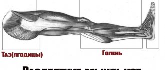
Gluteus maximus muscle, m. Gluteus maximus occupies almost the entire volume of the buttocks, so their shape largely depends on it. Beginning: gluteal surface of the ilium, dorsal surfaces of the sacrum and coccyx; insertion: gluteal tuberosity of the femur, iliotibial tract.
Main function:
ensure the movement of the hip joint, that is, straighten the torso, move the leg back.
Gluteus medius and minimus muscles
overlap the first and are located slightly above it. They are responsible for raising the leg to the side and also provide lifting of the buttocks.
Treatment
The piriformis muscle is located deep in the gluteal region. It is impossible to remove trigger points in it using classical massage techniques. The main treatment for problems caused by the piriformis muscle is manual therapy. In the presence of secondary disorders caused by pathology of the piriformis muscle, separate correction is required. Thus, neurovascular disorders in the affected limb, caused by compression of the vessels supplying the sciatic nerve, require the prescription of drugs that improve blood circulation. Depending on the severity of vascular disorders, drugs can be prescribed intravenously or in tablets. If the problems of the piriformis muscle are caused by a violation of its innervation, autogravitational traction of the spine is necessary.
As accompanying methods that accelerate the normalization of muscle tone and blood supply to the sciatic nerve, acupuncture, hirudotherapy, and some physiotherapeutic techniques are used. After pain relief, it is necessary to regularly perform special exercises for a long time to maintain normal muscle tone and form the correct pattern of movements.
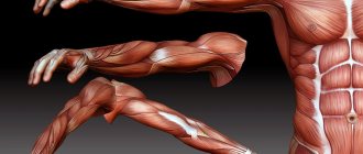
Muscles of the anterior thigh[edit | edit code]
Muscles of the anterior thigh and lower leg
Quadriceps (quadriceps)[edit | edit code]
Quadriceps femoris muscle, m. guadriceps femoris, the most powerful muscle in the human body. It occupies the entire front surface of the thigh. In front it is crossed obliquely by the sartorius muscle
.
Muscles of the anterior thigh
The muscle consists of four heads: rectus femoris, vastus internus, vastus externus and vastus medialis.
. Having different starting points far apart from each other, all the heads converge in the lower part of the thigh and form a common tendon.
Rectus femoris muscle
, m. rectus femoris, bipennate muscle, the longest of the four heads of the quadriceps muscle; located on the front surface of the thigh. The muscle begins as a tendon from the iliac spine and at the acetabulum. Heading down, the muscle expands and reaches the middle of the thigh, and then, gradually narrowing, turns into a powerful tendon. The latter fuses with the base of the patella and, passing along its anterior surface, reaches the tubercle of the tibia, where it ends.
Vastus internus muscle
, m. vastus medialis is a flat, wide, thick muscle. Located on the anteromedial surface of the thigh. Its anterior edge is somewhat covered by the rectus femoris muscle; inside it borders with the medial group of thigh muscles. The vastus internus muscle is partially covered by the oblique sartorius muscle. The muscle bundles, enveloping the anteromedial surface of the femur, are directed obliquely downward and forward. In the lower part of the thigh, the muscle part passes into the tendon part, which joins the rectus tendon.
Vastus externus muscle
, m. vastus lateralis is a flat, wide, thick muscle lying on the anterior outer surface of the thigh. Its lateral surface is partially covered by the tensor fasciae lata muscle; the anterior edge is covered by the rectus femoris muscle. The muscle starts from the trochanter of the femur. The muscle bundles, directed obliquely downward and forward, cover the anterolateral surface of the femur and in the lower part of the thigh pass into the tendon, intertwined with the tendon of the rectus muscle.
Vastus medialis
, m. vastus intermedius - the weakest of the four heads of the quadriceps femoris muscle; has the appearance of a flat, wide, relatively thin muscle. The muscle is located on the front surface of the femur, covered in front by the rectus femoris muscle. The muscle starts from the intertrochanteric line and the anterior surface of the femur within the upper three-quarters. Its bundles are directed vertically downwards and pass into a flat tendon. In the lower thigh, the tendon attaches to the rectus femoris tendon.
Main function:
the quadriceps femoris muscle extends the leg at the knee; in addition, m. The rectus femoris, being a biarticular muscle, takes part in hip flexion and forward tilt of the pelvis.
How can you relieve pain during exacerbation?
Mechanical overload of the muscle should be avoided. It is possible to correct asymmetry caused by inequality in the length of the lower limbs by using a special heel on shoes. You can correct the asymmetry caused by a decrease in the size of one half of the pelvis by using a pad under the buttock. It is important to restore movement in the sacroiliac joint.
Choosing the right sleeping position is of great importance. When sleeping on the side, the patient should place a pillow between the knees to support the leg. Frequent changes of sitting position are necessary; you can use a rocking chair. Stops should be arranged during long driving periods - for short walks.
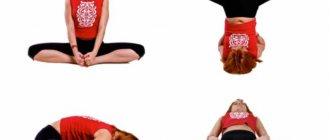
It is necessary to perform a set of exercises aimed at stretching the muscles, which include ischemic compression of trigger points. but it is very important to avoid compression of the nerves, so the exercises should be supervised by a specialist.
Hamstring muscles[edit | edit code]
Muscles of the back of the thigh and lower leg
Biceps femoris[edit | edit code]
Muscles of the back of the thigh
Biceps femoris, m. biceps femoris, placed closer to the anterior edge of the thigh. The muscle consists of two heads. Long, begins on the ischial tuberosity; short head, originating from the middle third of the lateral lip. Both heads, joined together, are attached to the head of the fibula.
Main function:
bending the knees and extending the torso together with the gluteus maximus muscle.
see also
: Hamstring muscles
The main causes of nagging pain in the thigh, buttock or groin
Finding the starting point of such a complaint often causes difficulties in differential diagnosis, since the causes of discomfort in these areas can be a consequence of a number of pathological conditions of the human musculoskeletal system. There are everyday reasons (lifestyle, work postures), acute diseases (injuries, exacerbations of chronic conditions), functional deviations in the functioning of the muscular corset.
Among the provocateurs of this pain syndrome are:
- excessive physical activity associated with work
- "sedentary work;
- problems with excess weight;
- the period of bearing a child.
Calf muscles[edit | edit code]
Triceps surae[edit | edit code]
Triceps surae muscle
m. triceps surae, forms the main mass of the roe elevation. It consists of two muscles - m. gastrocnemius, located superficially, and m. soleus, lying under it; both muscles below have one common tendon.
Calf muscle
m. gastrocnemius, starts from the femur behind both condyles with two heads that pass into the tendon, and continues into the massive Achilles tendon, which attaches to the posterior surface of the tubercle of the calcaneus.
Soleus muscle
, m. soleus, thick and fleshy. It lies under the calf muscle, occupying a large area on the bones of the lower leg. The line of its origin is located on the head and on the upper third of the posterior surface of the fibula and descends along the tibia almost to the border of the middle third of the tibia with the lower one. Fuses with the Achilles tendon. Posterior muscle group of the lower leg.
Plantaris muscle
, m. plantaris, originates above the lateral condyle of the femur and from the capsule of the knee joint, soon becomes a very long and thin tendon and inserts at the calcaneal tubercle. This muscle may be absent.
Main function:
produces flexion at the ankle joint both with the free leg and with support on the end of the foot. Since the line of pull of the muscle passes medially to the axis of the subtalar joint, it also causes adduction of the foot and supination. When standing, the triceps surae (especially the m. soleus) prevents the body from tipping forward at the ankle joint. The muscle has to work primarily when burdened by the weight of the whole body, and therefore it is strong and has a large physiological diameter; m. gastrocnemius, as a biarticular muscle, can also flex the knee when the lower leg and foot are strengthened.
The main muscles of the lower extremities and pelvis[edit | edit code]
| Name | Joints involved | Start | Place of attachment | Action | |
| Calf muscle | Ankle and knee | Condyles at the base of the femur | Posterior surface of the calcaneus | Ankle - plantar flexion of the foot (strong); Knee joint - shin flexion (weak) | |
| Soleus muscle | Ankle | Upper 2/3 of the posterior surface of the tibia and fibula. bones | Posterior surface of the calcaneus | Ankle - plantar flexion of the foot | |
| Quadriceps: Rectus femoris muscle | Hip and knee | Inferior anterior iliac spine of the pelvis | Patella and patellar ligament, attaches to the tibial tuberosity | Hip joint - hip flexion Knee joint - tibia extension | |
| Quadriceps: Vastus lateralis, intermedius, medial | Knee | Lateral, anterior and medial surfaces of the femur | Edge of the patella | Knee joint - tibia extension | |
| Hamstring muscle group: Short and long heads of the biceps femoris (lateral part); Semitendinosus and semimembranosus (medial part) (usually act as one muscle) | Hip and knee | The biceps femoris muscle - short head - on the back of the femur, the lower part of the line aspera and the intermuscular septum. The remaining heads begin from the ischial tuberosity of the pelvic bone | The biceps femoris muscle attaches to the head of the fibula and to the lateral condyle of the tibia. The semitendinosus/semimembranosus muscles attach to the medial condyle of the tibia | Hip joint - hip extension; Knee joint - flexion of the tibia; Biceps femoris ext. outward rotation of the thigh and lower leg (supination). The semitendinosus/semimembranosus muscles rotate the femur and tibia inward (pronation) | |
| Adductor muscle group: Pectineus muscle, short. drive unit. muscle, length drive, muscle, pain drive, muscle, gracilis (act as one muscle) | Hip (gracilis femoris muscle also crosses the knee joint) | Pubic and ischial bones of the pelvis | Int. over, thighs bones, to the lesser trochanter of the femurs. bones, to the rough line on the back of the thighs. bones; and a large drive, m. to the medial epicondyle of the femurs. bones; Thin m. thigh to tibia. bones | Main function: hip adduction | |
| Tensor fascia lata | Hip | Superior anterior iliac spine | Weaves into the iliotibial tract | Abduction, hip flexion and participation in hip internal rotation | |
| Gluteus maximus muscle | Hip | Rear over the iliac crest, sacrum and fascia of the back in the region. explanatory vertebrae | Partially passes into the iliotibial tract into the fascia lata | Extension, external rotation of the hip | |
| Gluteus medius and minimus muscles | Hip | Outer surface of the ilium | Greater trochanter of the femur | Hip abduction, involved in turning the hip outward and inward | |
| Iliopsoas muscle | Hip | Int. over, ilium, bases, sacrum; side. on top, ate breast and waist. vertebrae | Lesser trochanter of the femur | Hip flexion | |
| Deep rotator muscles: Piriformis, Gemini (superior and inferior), Obturator (external and internal), Quadratus femoris (located under the gluteus maximus) | Hip | Anterior surface of the sacrum, posterior surface of the ischial tuberosity and edges of the obturator foramina | Apex and trochanteric fossa of the greater trochanter of the femur | Hip abduction and external rotation |
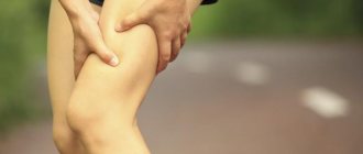
Symptoms of trauma.
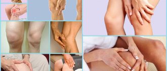
A characteristic and primary symptom for all types of tendon injuries is the appearance of sudden severe pain. The following signs are added to it:
- redness and swelling in the area of injury
- increased sensitivity at the site of injury
- muscle cramps
- hemorrhage
- characteristic crunching sound at the time of injury
- limited movement of the injured limb
- complete ruptures are characterized by the appearance of dips when walking
Muscles around the toe joints[edit | edit code]
Foot muscles
- Extensor hallucis brevis
- Extensor hallucis longus
- Extensor toes brevis
- Extensor toe longus
- Flexor hallucis brevis
- Flexor hallucis longus
- Flexor toe brevis
- Flexor toe longus
- Quadratus plantaris muscle
- Flexor of the little toe brevis
- Dorsal interosseous muscles of the foot
- Abductor hallux muscle
- Abductor of the little toe muscle
- Adductor hallucis muscle
- Plantar interosseous muscles
- Vermiform muscles of the foot
Fabric channels
The femoral triangle is what is located in the upper region of the limb. It is formed by adjacent muscles and ligaments. It contains nerves and blood vessels. The connective tissue membrane consists of two plates, between which a channel of the same name is formed, consisting of a pair of holes and three walls.
Catheterization is performed in this place, digital pressure is used to stop bleeding.
The thigh muscles are among the strongest developed in the human body. They hold the torso in an upright position, control the hip and knee joints, and are used during various activities, and even at rest. Their proper functioning is extremely important, and any person should know the principle of operation.
For ease of understanding, they are divided into 3 sections. Let's look at each of the points in more detail, and also look at the images.
Functions
1 The largest muscle group during training that burns a large number of calories, increasing the metabolic rate.
2. They add muscle volume and mass during development.
3. Develop performance and strength by performing various strength exercises.
4. Strengthen muscles, making the corset more reliable. Testosterone and muscle building hormones are produced when performing various leg exercises.
5. The muscles of the arms, shoulders and back (upper body) develop.
6. They build a body that is more proportional in appearance (if you compare the top and bottom).
7. Develop a more powerful connection between muscles and the brain, that is, neuromuscular.
8. Strengthens concentration and willpower.
9. Strength training helps prevent certain diseases, osteoporosis and arthritis.
To avoid suffering from these joint diseases, you need to perform leg exercises with a moderate load.
10. Strong legs prevent getting any blows and various sprains.
11. Training has a good effect on the cardiovascular system and the functioning of the heart itself.
12. Beautiful, slender legs do not need to be hidden under clothes, so you can wear looser clothes.
13. Buttocks with good volume and elasticity will attract the attention and affection of men. After all, it is physiologically inherent that this determines the fertility and health of a woman.
Exercises for each muscle group
In bodybuilding and fitness, it is very difficult to load the legs so that one individual muscle works, therefore all movements are classified into groups. Each exercise corresponds to the main function of a particular area.
Buttocks
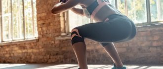
- Squats with a barbell (only with a deep squat, below the parallel of the thigh with the floor);
- Lunges forward;
- Gluteal bridge;
- Leg press (only with high feet on the platform).
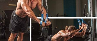
Quadriceps
- Squats;
- Leg press;
- Leg extensions while sitting in a machine;
- Classic lunges.
Biceps hamstrings
- Romanian deadlift;
- Lifting the body in a simulator (hyperextension);
- Lying leg curls in a machine.
Adductor muscles
- Squats with wide legs (sumo);
- Bringing the legs in a crossover;
- Leg abduction in the simulator.
Caviar
- Standing and sitting calf raises (with free weights or in a machine);
- Exercise "Donkey".
Diagnostics
The examination is usually carried out by orthopedic traumatologists. In case of purulent processes, patients are examined by a surgeon, in case of neurological pathology - by a neurologist. At the initial stage, the doctor finds out the history of life and illness, and conducts an external examination. The following additional studies are prescribed:
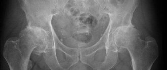
- X-ray of the hip.
A basic diagnostic method that allows you to identify all major bone pathologies: fractures, cancer, osteomyelitis, periostitis. In case of lesions of soft tissue structures, it is performed to exclude bone lesions and conduct differential diagnosis. - CT scan of bone.
It is used at the final stage of the examination to clarify the results of radiography, planning conservative therapy or surgical intervention. Makes it possible to accurately determine the size, configuration and structural features of the pathological focus in bone tissue. - Ultrasound.
Sonography of soft tissues in myositis and tendonitis reveals areas of inflammation or degeneration. Ultrasound scanning of the veins of the lower extremities is effective in assessing the condition of the veins and lymphatic vessels in patients with phlebitis and lymphangitis, and in identifying blood clots in thrombophlebitis. - MRI of soft tissues.
Recommended for ambiguous results of other diagnostic procedures. It is carried out to determine the localization of deep hematomas, confirm degenerative or inflammatory changes in tendinitis and myositis. - Electrophysiological studies.
For patients with pain of neurological origin, ENMG, ENG or EMG are indicated to clarify the level of nerve damage and study the condition of nerve and muscle tissue. - Lab tests
. They are performed to determine the severity of the inflammatory process in purulent diseases, assess the condition of organs and systems in malignant neoplasms.