Identification of antagonist muscles
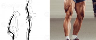
Most of the muscles of the torso, arms and legs are located in opposite pairs.
This means that when one muscle makes a contraction movement, such as the biceps, that muscle acts as an agonist for the other muscle during the exercise. Then the opposite muscle, in this case the triceps, is called the antagonist muscle. When we talk about agonist and antagonist muscles, we are talking about the role that certain muscles play in your range of motion. Knowing how these muscles are structured and the role they play in exercise, especially when it comes to concentric, eccentric, and isometric contractions, can help you take a more targeted approach to your training, which will more effectively move you closer to your fitness goals. In fact, using opposing muscle groups is one of the most popular strength training methods because you eliminate rest periods, which can save you time. Since antagonistic muscles work in synergy, it is important that both muscles are trained equally and receive the same load. This can be done sequentially, performing a series of exercises aimed at developing one muscle, and then moving on to training its antagonist, thus alternating training the opposite muscles.
The training program for antagonistic muscles has been incredibly popular among athletes for decades. This is nothing new, Arnold Schwarzenegger was the champion of antagonistic supersets back in the 1970s. In the world of strength sports, antagonistic supersets were popularized by Charles Poliquin and Ian King in the early 1990s.
This training approach has a number of justified advantages, and with proper use of the acquired knowledge about the principles of work of antagonist muscles, it allows you to reach a qualitatively new level of sports achievements. Before jumping directly into the training program, let's summarize its key benefits.
About muscle tissue and the nuances associated with it
Greetings, dear colleagues!
Much of a fitness trainer's job involves muscle tissue, and today I'm going to give you some general information about it from an academic anatomy perspective. There is a high probability that you will make a number of discoveries for yourself.
This will help you better understand both basic fitness training if you are just planning to become a trainer, and better master the material of courses for existing fitness instructors.
Myology is a branch of anatomy devoted to the study of muscles.
Types of muscle tissue:
- smooth (internal organs, involuntary contraction, unconsciously controlled);
— striated:
- cardiac (heart, automatic, involuntary contraction, unconsciously controlled)
- skeletal (with the help of tendons it is attached to the bones (muscles) + as part of the gastrointestinal tract, eyes, etc., voluntary contraction, consciously controlled)
Skeletal muscle is an organ with a characteristic shape and structure, built from bundles of striated muscle fibers interconnected by loose connective tissue (endomysium, perimysium and epimysium) and covered on the outside with its own fascia.
There are about 600 skeletal muscles in humans; total weight – up to 40% of body weight.
In the muscle there are:
- head (caput) - the initial part;
- body (corpus) - middle part;
- tail (cauda) - the final part.
The length of the muscle determines the degree of amplitude it can provide.
Each muscle has an origin and an insertion point.
The origin of the muscle, as a rule, is a fixed point (punctum fixum), and the place of attachment is movable (punctum mobile).
For example, the subclavian muscle has its origin at the first rib and its insertion point at the clavicle.
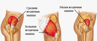
The gluteus minimus originates from the ilium and attaches to the greater trochanter of the femur.
But there are exceptions: depending on the function performed, attachment may be the beginning. For example, when the pectoral muscles become auxiliary respiratory muscles.
The connections between tendon and bone are called enthesis (Latin: enthesis) or insertions (Latin: insercio).
Moreover, in children, on average, up to 13-16 years of age, “rough” places of muscle attachment, which are located near the main areas of bone growth (calcaneal tubercle, greater trochanter of the femur, greater tubercle of the shoulder, tibial tuberosity, etc.), are called apophyses .
Functions of skeletal muscles:
- change the position of the human body and its parts;
- participate in the formation of cavity walls;
- are part of the organs (tongue, esophagus, organs of hearing and vision);
- participate in breathing and swallowing;
- provide physiological functions (childbirth, urination, defecation);
- blood and lymph flow;
- participate in thermoregulation (heat production - due to contraction);
- muscular-articular sense (proprioception, large receptor field).
In each muscle there are main and auxiliary apparatuses.
main office:
- active (contracting) part - muscle belly, venter;
- passive part (attached to the bone) – tendon, tendo.
The broad tendon is called an aponeurosis .
auxiliary apparatus are formations that facilitate the work of muscles:
fascia, fascia , is connective tissue that covers individual muscles and muscle groups with a sheath.
Functions:
- separation of muscles from each other;
- reducing friction between adjacent muscles;
- support for the contracting muscle;
- directed muscle contraction;
- the origin or insertion of other muscles;
- formation of sheaths for neurovascular bundles;
- isolation of inflammation;
- ways of spreading pus and blood;
- "case" anesthesia;
- obstruction of the collapse of veins passing through the fascia.
synovial tendon sheath , vagina synovialis tendinis , are synovial membranes in the form of sheaths around the tendons to reduce friction between the tendons.
For example, tendinitis of the long head of the biceps often occurs precisely due to microdamage to the tendon sheath.
synovial bursae, bursae synoviales , are closed cavities filled with synovium, located under the muscles and tendons at the points of their contact with the bone in the joint area to reduce friction of the muscle/tendon on adjacent bones.
They can communicate with the joint cavity.
sesamoid bones, ossa sesamoidea , are bones located in the thickness of muscle tendons near the point of attachment in order to strengthen the joints and increase the leverage of the muscle (for example, the patella, patella).
muscle retinaculum, retinaculum , - thickened ligaments or sections of fascia that spread between bony protrusions above the muscle tendons. At the same time, they form channels together with the bones (examples are the wrist and the back of the foot).
Latin nomenclature and basic principles of muscle classification.
direction - orientation of muscle bundles relative to the sagittal plane:
- rectus - straight;
- transversus - transverse;
- obliquus - oblique;
comparative muscle size:
- maximus - the largest (example - m. gluteus maximus, gluteus maximus muscle);
- minimus - the smallest (example - m. gluteus minimus, gluteus minimus);
- longus - long;
- brevis - short;
- latissimus - widest;
- longissimus - longest;
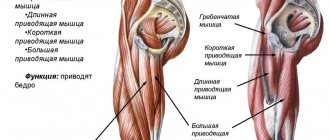
- magnus - large (example - m. adductor magnus, adductor magnus muscle);
- major - larger (example - m. pectoralis major, pectoralis major muscle);
- minor - smaller (example - m. pectoralis minor, pectoralis minor muscle);
- vastus - wide (example - m. vastus medialis, vastus medialis muscle (head of the quadriceps);
comparative muscle shape:
- deltoideus - deltoid;
- trapezius - trapezoidal;
- serratus - jagged;
- rhomboideus - rhomboid;
- orbicularis - circular (example - mm. orbiculares oris et oculi, circular muscles of the mouth and eye)
- pectineus - comb;
- piriformis - pear-shaped;
- quadratus - square;
- gracilis - thin, delicate;
function:
- flexor - bends, reduces the articular angle;
- extensor - extends, increases the articular angle;
- adductor - leads, moves the bone away from the midline;
- abductor - abducts, moves the bone towards the midline;
- levator - lifts up a part of the body;
- depressor - lowers a part of the body;
- supinator - external rotation (turns the palmar surface forward);
- pronator - internal rotation, turns over (palm surface back);
- sphincter - reduces the lumen of the hole;
- tensor - strains;
number of origins - the number of tendons (abdomens, “heads”) at the point of origin of the muscle:
- biceps - two-headed;
- triceps - three-headed;
- quadriceps - four-headed;
localization - the structure around which the muscle is located (example: m. temporalis - temporal muscle);
places of attachment - the place of origin and attachment of the muscle (example: m. sternocleidomastoideus - sternocleidomastoid muscle), etc.
Synergists are muscles that perform the same function.
Antagonists - perform the opposite function.
Of the two antagonist muscles, the one that carries out a given movement (that is, performs the main task) is called agonist , and the other is called antagonist.
Classification by topography:
- superficial muscles;
- deep muscles.
Classification by body area:
- facial muscles and chewing muscles;
- neck muscles;
- muscles of the trunk (back, chest, abdomen);
- muscles of the upper limb;
- muscles of the lower limb;
- muscles of the perineum (pelvis and genitourinary diaphragm).
Classification in relation to joints:
- single-joint (act on 1 joint);
- two-joint;
- multi-joint;
- do not affect joints (facial, perineum).
When discussing any skeletal muscle, it is recommended to follow a simple framework:
1. name (Russian and Latin);
2. points of origin and attachment (do not confuse them!);
3. functions.
What conclusions can be drawn?
1. ligament (ligamentum) connects bones, and tendon (tendo) connects the muscle belly and bone;
2. enthesopathy = insertionitis - this is a special case of tendinopathies;
3. for children, the types of apophyseal pathology are apophyseal fractures and one of the types of osteochondropathy (Osgood-Schlatter disease, Schinz disease);
4. fascia is an auxiliary component of the muscle; it is technically impossible to train them separately;
5. Since some muscles, depending on their function and individual characteristics, can change their points of origin and insertion, in the case of muscle correction it is necessary to ensure through several repetitions of manual directional examination that it is in this direction that the thrust should be created.
You can better study muscle balance, diagnostic tests and methods of work in the Prehab course ( details >>> )
Author: neurologist Nikolai Votchitsev
Fitness training for trainers.
For fitness trainers looking for courses that can increase their income, make them even better, and ensure they never have to worry about finding a job, we recommend checking out the following distance courses:
Prehab is a distance course for fitness trainers who want to fully understand the topic of working with muscle balance and improving the movement of their charges.
The basic personal trainer course is for those instructors who want to supplement their knowledge with fundamental information. An incredibly large amount of useful material that will take you to the next level.
Power is an online course for fitness trainers who are passionate about functional and strength training and want to better understand the principles of these areas, as well as increase their income.
Body Architecture - Dmitry Gorkovsky's original course with face-to-face practical days and distance theory for fitness trainers, massage therapists and doctors.
Pregnant - training methods for working with pregnant women and recovery after childbirth.
What are these advantages?
Effective use of training time. For busy people who want to get the most out of their time at the gym, this is invaluable. Maintaining muscle balance. Our muscles are connected by fascial systems, networks of soft tissue that surround the muscles and allow them to move in conjunction with each other. If there is one tight or underdeveloped muscle in the fascial system, it can limit the mobility and strength of the rest of the system and lead to excessive tension in other areas. Increasing the intensity of training and, as a result, strength indicators. As already mentioned, while one muscle is subject to increased tension, the antagonist muscle at this very time is at rest or in a slight static tension. This allows you to better alternately work large muscle groups, for example, the muscles of the back and chest.
Synergy and synergists
The most important aspect in understanding how muscles function to create joint movement is synergy. Synergy means that two or more units work together to achieve a result. Working together, the entire result will be greater than the sum of the individual effects of the agents involved. Even the simplest joint movement requires muscles to work together in this synergistic or cooperative manner. When a group of organs work together to optimally perform a given motor task, it is called muscle synergy.
Typically, contractile organs that are directly involved in creating a certain joint movement are called agonists and those that are indirectly associated with some other role are called synergists. However, even if a muscle acts directly to move a joint, adding its own torque, it can still be correctly called a "synergist." Other muscles, such as stabilizers, neutralizers, and fixators, which aid movement by counteracting unwanted movement or helping stabilize a joint are also synergistic.
Synergists and antagonists: an interesting explanation with examples awaits on the lines below.
Principles of competent construction of a training program
When starting to develop a program for training antagonistic muscles, do not forget about the simple rule - “more” does not mean “better”. Excessive zeal at the start and overtraining are the most common reasons for lack of progress, and often injuries. All this can weaken motivation. To build a competent training program, you should understand that muscle strength is not built up during training, but during rest . During training, muscle microtraumas occur, while during rest, all the body’s forces are directed toward restoring the damage received. It is during recovery that physiological adaptation to loads occurs - the muscle becomes stronger and more powerful to withstand the upcoming loads. Therefore it is important:
- observe the time intervals allotted for recovery;
- correctly dose the load.
General overview of the triceps brachii.
The triceps brachii muscle is a muscle of the posterior group of shoulder muscles. As with the biceps brachii, the triceps in humans was modified by the importance of the throws. The triceps brachii muscle is part of the trigger system in this slingshot concept. In primates, the triceps are more powerful/stronger, but, as with the biceps, less functional.
Biologists note that the accuracy and throwing power of primates is very much inferior to these same characteristics in humans. The triceps brachii muscle plays an important role in this. In addition to important functions such as throwing and using objects, the triceps brachii is involved in a huge number of everyday movements - from getting up from a table to opening a door.
Like the biceps, the triceps brachii muscle shapes the appearance of the shoulder, giving it most of its volume since it is larger than the biceps muscle.
Triceps origin points.
- long head (caput longum) - subarticular tubercle of the scapula (tuberculum infraglenoidale scapulae);
- medial head (caput mediale) - the posterior surface of the humerus below the groove of the radial nerve (sulcus n. radialis), lateral and medial intermuscular septa;
- lateral head (caput laterale) - the posterolateral surface of the humerus above the groove (sulcus n. radialis), lateral and medial intermuscular septa).
Attachment of the triceps brachii muscle.
All three heads in the lower half of the shoulder converge into a powerful abdomen, which is attached to:
- posterior surface of the olecranon (olecranon) of the ulna;
- elbow joint capsule;
- own fascia of the forearm (fascia antebrachii).
Triceps fiber direction.
The fibers are directed downward and converge in a semicircle in a common abdomen.
Blood supply, innervation and lymphatic drainage.
Arterial blood is delivered by the branches of the deep brachial artery (a. profunda brachii). Venous blood flows through the named veins.
Innervation is carried out by the radial nerve (n. radialis, myotome C7−8), which is a branch of the brachial plexus. However, according to some studies, the medial head is innervated by the ulnar nerve, and the long head by the axillary nerve. Lymphatic drainage is carried out into the axillary lymph nodes (nodi axillares) and then into the subclavian trunk (truncus subclavius).
CONTENT
- 1 Types 1.1 Skeletal muscle
- 1.2 Smooth muscle
- 1.3 Cardiac muscle
- 2.1 Agonists and antagonists
- 3.1 Insertion and origin 3.1.1 Origin
- 4.1 Hypertrophy and atrophy
Types[edit]
There are three types of muscle tissue in the human body: skeletal, smooth and cardiac.
Skeletal muscle[edit]
Skeletal striated muscle, or "voluntary muscle", is primarily connected to bone by tendons. Skeletal muscles provide movement to the bones of the human skeleton and maintain posture. [1]
Smooth muscle[edit]
Smooth muscle tissue is found in those parts of the body where it transmits action without conscious intention. Most of this type of muscle tissue is found in the digestive and urinary systems, where it acts to propel food, chyme and feces in the former and urine in the latter. Other places where smooth muscle can be found are the uterus, where it helps facilitate childbirth, and the eye, where the pupillary sphincter controls pupil size. [2]
Cardiac muscle[edit]
Cardiac muscle is specific to the heart. It also moves involuntarily and, in addition, self-excites, contracting without external stimuli. [3]