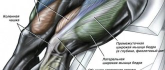
Skeletal muscle of the thigh
| Adductor longus muscle | |
| Adductor longus and nearby muscles | |
| Structures surrounding the right hip joint. (Adductor longus muscle at top right.) | |
| Details | |
| Source | pubic body just below the pubic ridge |
| Insert | middle third linea aspera |
| Artery | deep femoral artery |
| Nerve | anterior branch of the obturator nerve |
| Actions | hip adduction, hip flexion |
| Identifiers | |
| Latin | Musculus adductor longus |
| TA98 | A04.7.02.026 |
| TA2 | 2628 |
| F.M.A. | 22441 |
| Anatomical terms of the muscle [edit in Wikidata] | |
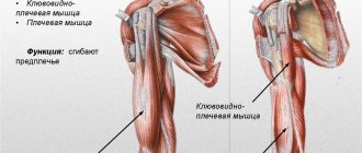
In the human body, the adductor Longus
is a skeletal muscle found in the thigh. One of the adductor muscles of the thigh, its main function is to adduct the femur and is innervated by the obturator nerve. It forms the medial wall of the femoral triangle.
Links[edit]
- ^ a b c d e Platzer, Werner (2004). Color Atlas of Human Anatomy, Vol. 1, musculoskeletal system (5th ed.). Time. item 242.
- Jump up
↑ Wilson, Erasmus (1851). Anatomist's vade mecum: a system of human anatomy. John Churchill. paragraph 260. - Saladin, Kenneth S. Anatomy and Physiology: Unity of Form and Function. 5th ed. Boston: McGraw-Hill, 2009.
- Aatif M. Hussain (2008). A Practical Approach to Neurophysiological Intraoperative Monitoring. Demos Medical Publishing. paragraph 23.
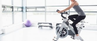
Sample training program for the hip adductors
We recommend combining training of the adductors with training of other muscles of the lower body.
Example:
- Warm up on a jump rope or treadmill for 5 minutes;
- Squats (4/6-8);
- “Wide” leg press in the simulator (3/10-12);
- Forward lunges with kettlebells (3/10-12);
- Leg abduction in the simulator (3/10-12);
- Calf raises in a vertical machine (3/15-20).
The considered example of an activity is more suitable for men. Girls can adapt the training to suit themselves, for example, replacing classic squats with a plie variation. After working out your lower body, we recommend doing some simple stretching.
Additional images[edit]
- Right femur. External surface.
- Muscles of the iliacus and anterior femoral regions.
- Deep muscles of the medial femoral region.
- Left femoral triangle.
- Femoral artery.
- Lumbar plexus and its branches.
- Transverse section of the thigh.
- Adductor longus muscle
- Adductor longus muscle
- Adductor longus muscle
- Adductor longus muscle
- Adductor longus muscle
- Adductor longus muscle
- Adductor longus muscle
- Adductor longus muscle
- Adductor longus muscle
- Thigh muscles. Cross section.
- Thigh muscles. Front views.
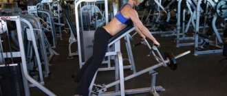
Basic quadriceps exercises.
- Squats.
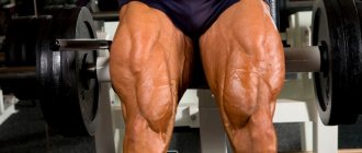
Don't be afraid of hardware. Many girls are afraid of too swayed legs. In fact, achieving them is not so easy - this requires high-repetition exercises with progressive load.
The barbell squat is a universal exercise that allows you to strengthen not only your quadriceps, but also your buttocks, back and abs. It all depends on following the correct technique for performing the exercise.
To avoid injury, make sure that the barbell rests on the trapezius muscle and not on the neck. It is best to hold the barbell with a narrow grip. In this case, the chest and back should be level, without bending. Place your feet shoulder-width apart and point your toes slightly out to the sides.
The squat movement should begin by moving the pelvis back. The knees should remain as fixed as possible: not bend back, but also not move forward.
The bottom point of the squat is parallel to the thighs with the floor. If you go lower, the load on the quadriceps will become significantly less. The gluteal muscles will already be included in the work.
When straightening your knees, make sure that they do not straighten all the way.
The feet should be firmly on the surface, the heel should not come off it. To control the position of your back, try to look forward and feel the work of the target muscle.
For a home workout, dumbbells are quite suitable instead of a barbell. Hold them in your hands, downwards.
- Front squats with a barbell.
Due to the non-standard placement of the barbell, its weight should be lower than with the classic version of the exercise. For complete beginners, it is better to try doing squats in a Smith machine. This exercise works especially well on the medial head of the quadriceps.
Place the barbell on your front deltoids. At the same time, the grip of the barbell is narrow with the arms crossed. Try not to arch your back and squat until it is parallel to the floor.
- Leg press.
Another exercise that can be done in the gym. To engage the quadriceps muscles, place your feet shoulder-width apart.
Unlike the squat, the weights used in the machine can be significantly heavier.
To begin, take the correct body position in the machine: your back and head are pressed tightly against the back of the machine. Bend your knees until you form a 90-degree angle.
When you reach the top point in the machine, your knees should remain slightly bent.
There are variations for the leg press. It can be performed with both legs at the same time, or with one leg at a time.
- Squats in the Hack machine.
This type of squat is an inverted form of the previous exercise. It’s just that in this case, the upward movement is made by the body, not the legs.
After you sit on the platform, place your feet shoulder-width apart, and rest your shoulders on special pillows. Lower yourself down, bending your knees until they are parallel to the floor. Repeat the upward pushing movements of your body.
Make sure your heels are firmly planted on the platform and do not move away from it. Your back should be pressed into the back of the machine.
- Leg extension in the simulator.
This exercise thoroughly works the quadriceps or quadriceps muscle.
To perform it, you need to sit in a special simulator and set the required height of the rollers, which you will push upside down. As you exhale, push your quadriceps legs up, and as you inhale, gently lower them down.
- Lunges.
For those who cannot go to the gym, you can always find an alternative. In order to load the quadriceps, you can perform various squats. For the greatest load on your shoulders, you can put additional weight - a barbell, a sandbag, or pick up dumbbells. Lunges can be done in one place or while walking. It all depends on your territorial capabilities.
As with the previous exercises, the angle at the knee should be 90 degrees. Don't forget to watch your breathing. Lower your body as you inhale, lift it as you exhale. Lunges can also be performed alternately on both legs or on the same limb.
- "Pistol."
Although this exercise seems accessible, due to the fact that you do not need additional sports equipment, it is not at all easy to perform. If in other exercises it is possible to incorrectly redistribute the load on non-target muscles, then a squat on one leg practically eliminates this. The main work here is performed by the quadriceps.
Abstract
The present study investigated the longitudinal growth of the vastus lateralis muscle using four eccentric exercise protocols with different mechanical stimuli by modifying the load magnitude, lengthening velocity and muscle length at which the load was applied.
Thirty-one participants voluntarily participated in this study in two experimental and one control group. The first experimental group (N=10) exercised the knee extensors of one leg at 65% (low load magnitude) of the maximum isometric voluntary contraction (MVC) and the second leg at 100% MVC (high load magnitude) with 90 deg/ s angular velocity, from 25 to 100 deg knee angle. The second experimental group (N=10) exercised one leg at 100% MVC, 90 deg/s, from 25 to 65 deg knee angle (short muscle length). The other leg was exercised at 100% MVC, 240 deg/s angular velocity (high muscle lengthening velocity) from 25 to 100 deg.
In the pre- and post-intervention measurements, we examined the fascicle length of the vastus lateralis at rest and the moment–angle relationship of the knee extensors. After 10 weeks of intervention, we found a significant increase (~14%) of vastus lateralis fascicle length compared with the control group, yet only in the leg that was exercised with high lengthening velocity.
The findings provide evidence that not every eccentric loading causes an increase in fascicle length and that the lengthening velocity of the fascicles during the eccentric loading, particularly in the phase where the knee joint moment decreases (ie deactivation of the muscle), seems to be an important factor for longitudinal muscle growth.
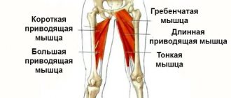
Medial group
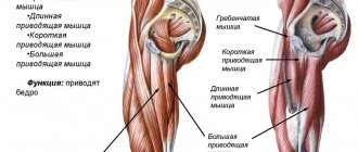
The pectineus muscle (m. pectineus) (Fig. 90, 129, 130, 132) flexes and adducts the thigh, rotating it outward. The flat muscle is quadrangular in shape, originates on the crest and superior ramus of the pubis, and is inserted on the medial lip of the linea aspera of the femur below the lesser trochanter.
The thin muscle (m. gracilis) (Fig. 90, 129, 130, 132, 134, 145) adducts the thigh and takes part in flexing the tibia, turning the leg inward. The long, flat muscle is located just under the skin. Its point of origin is on the lower branch of the pubic bone, and its attachment point is on the tuberosity of the tibia. The gracilis tendon fuses with the sartorius and semitendinosus tendons and the fascia of the leg to form the superficial pes anserine. The so-called goose bursa (bursa anserina) is also located here.
| Rice. 133. Muscles of the pelvis and thigh, side view 1 - latissimus dorsi; 2 - external oblique abdominal muscle; 3 - gluteus medius muscle; 4 - gluteus maximus muscle; 5 - sartorius muscle; 6 - muscle that tightens the fascia lata of the thigh; 7 - iliotibial tract; 8 - the longest rectus femoris muscle; 9 - biceps femoris muscle: a) long head, b) short head; 10 - vastus lateralis muscle; 11 - calf muscle |
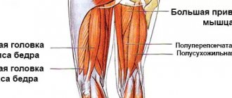
| Rice. 134. Muscles of the pelvis and thigh, rear view 1 - gluteus maximus; 2 - adductor magnus; 3 - iliotibial tract; 4 - tendon jumper of the semitendinosus muscle; 5 - semitendinosus muscle; 6 - biceps femoris muscle; 7 - thin muscle; 8 - semimembranosus muscle; 9 - sartorius muscle; 10 - plantaris muscle; 11 - gastrocnemius muscle a) medial head, b) lateral head |
The long adductor muscle (m. adductor longus) (Fig. 90, 129, 130, 132) adducts the thigh and takes part in its flexion and outward rotation. This is a flat muscle, shaped like an irregular triangle and located on the anteromedial surface of the thigh. It starts from the superior ramus of the pubis and inserts on the middle third of the medial lip of the linea aspera of the femur.
The short adductor muscle (m. adductor brevis) (Fig. 131) adducts the thigh and takes part in its flexion and outward rotation. It is a triangular-shaped muscle that originates on the anterior surface of the inferior ramus of the pubis, lateral to the gracilis muscle, and is inserted on the upper third of the medial lip of the linea aspera of the femur.
The large adductor muscle (m. adductor magnus) (Fig. 129, 130, 131, 132, 134) adducts the thigh, partly rotating it outward. Thick, wide, the most powerful muscle of this group, located deeper than the other adductor muscles. Its point of origin is located on the ischial tuberosity, as well as on the branch of the ischium and the lower branch of the pubic bone. The attachment site is located on the medial lip of the linea aspera and the medial epicondyle of the femur. Several holes are formed in the muscle bundles, allowing blood vessels to pass through. The largest of them is called the tendon opening (hiatus tendineus). Above it is a fascial plate, and between it and the muscle a triangular-shaped space is formed, called the adductor canal (canalis adductorius) (Fig. 131). The femoral vein, artery and hidden nerve of the lower limb pass through it.
Tips for maximum efficiency
- Before performing the above exercises, it is worth doing a WARM-UP. We pay special attention to the root joints and lower back.
- In each exercise we perform 4 approaches, 10-12 repetitions.
- For beginners, it is best to work without weight until you master the technique. Afterwards, slowly add weights.
- When performing, you need to feel the tension of the adductor muscles. Movements should be smooth.
- If your adductors are a lagging muscle group, then you should train them at the beginning of your workout after squats.
- Choose 2-3 exercises in which you best feel the desired muscle groups and take them into service.
Now you are armed with exercises that can pump up the muscles of the inner thigh. Stick to the technique and you can achieve excellent results. If you train at home, consider purchasing dumbbells and a resistance band. With them, the path to massive thighs will go much faster.
Good luck to everyone in your training!
Back group
The biceps femoris muscle (m. biceps femoris) (Fig. 133, 134, 145) extends the thigh and flexes the lower leg. In a bent position, rotates the lower leg outward. It runs along the lateral edge of the upper thigh. The muscle has one belly and two heads. The long head (caput longum) starts from the ischial tuberosity, the short head (caput breve) - on the lower part of the lateral lip of the linea aspera of the femur. The abdomen ends in a long narrow tendon, the attachment point of which is on the head of the fibula. Some of the bundles are woven into the fascia of the leg. Near the point of origin of the long muscle is the superior bursa of the biceps femoris muscle (bursa m. bicipitis femoris superior). In the area of the tendon there is the lower subtendinous bursa of the biceps femoris muscle (bursa subtendinea m. bicipitis femoris inferior).
The semitendinosus muscle (m. semitendinosus) (Fig. 130, 132, 134, 145) extends the thigh, bends the lower leg, rotating it inward in a bent position, and also takes part in straightening the torso. The muscle is long and thin, partially covered by the gluteus maximus muscle, sometimes interrupted by a tendon bridge (intersectio tendinea) (Fig. 134). Its origin point is located on the ischial tuberosity, and its attachment point is on the medial surface of the tibial tuberosity. Individual bundles of muscles are woven into the fascia of the leg, taking part in the formation of the crow's foot.
The semimembranosus muscle (m. semimembranosus) (Fig. 130, 132, 134, 145) extends the thigh and bends the tibia, rotating it inward. It runs along the medial edge of the posterior surface of the thigh and is partially covered by the semitendinosus muscle. The muscle starts from the ischial tuberosity and attaches to the edge of the medial condyle of the tibia.
The tendon is divided into three bundles, forming a deep pes anserine. The external bundle passes into the popliteal fascia, into the posterior ligament of the knee joint.
At the site where the tendon divides into separate bundles, the synovial bursa of the semimembranosus muscle (bursa m. semimembranosi) is located.