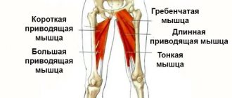
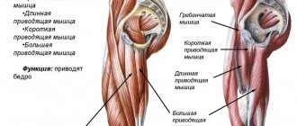
Adductor muscles (longus, brevis and adductor magnus muscles)[edit | edit code]
Adductor muscles (longus, brevis and adductor magnus)
Home[edit | edit code]
- Adductor longus
: superior ramus of the pubis - Adductor brevis
: inferior ramus of the pubis - Adductor magnus
: inferior ramus of the pubis, ramus of the ischium to the ischial tuberosity
Attachment[edit | edit code]
- Adductor longus
: middle third of the medial labrum of the linea aspera of the femur - Adductor brevis
: upper third of the medial labrum of the linea aspera of the femur - Adductor magnus
: medial labrum of the linea aspera of the femur, on the medial epicondyle of the femur
Innervation[edit | edit code]
- Obturator nerve L2-L4
When to expect the effect
If workouts that strengthen muscle tone are carried out with a large number of repetitions, and the weight of the dumbbells is light, then the result can be seen within 2-3 weeks. Such training does not bring much muscle fatigue and pain. It is advisable to perform exercises on other muscle groups the next day.
Then, with regular training, all muscles will be tense and ready for work, and constant tone will lead not only to strengthening muscle tissue, but also to weight loss. It should be remembered that the effect of toning exercises on the adductor muscles of the thigh is short-term.
After canceling training, the muscles “deflate”. Anatomically, the muscle structures of the thigh have a deep location and therefore many people forget about them, being carried away by pumping up the abdominals and buttocks. If the internal muscles are not active and pumped up enough, then the target muscles will be weakly loaded.
Participation in sports[edit | edit code]
As hip adductors, the adductors perform dynamic work in sports such as water polo, speed skating, and all team sports (acceleration when changing direction) and static work in luge, triple jumps and fencing. These muscles perform dynamic and static work in all running and walking disciplines, equestrian sports, alpine skiing and artistic gymnastics. As external rotators, they are involved in the execution of movements in figure skating, gymnastics, discus throwing and contact sports. The adductor longus and brevis muscles are involved in hip flexion during walking and running on the side of the carried leg, and the adductor magnus muscle participates in the work of the gluteus maximus muscle during extension in the stance phase.
| Kind of sport | Movement/hold | Function | Load | Types of abbreviations |
| Water polo | Walking in water | Hip adduction | Strength endurance | Dynamic concentric |
| Team sports | Acceleration when changing direction | Hip adduction | Fast, explosive | Dynamic concentric |
| Triple jumps | Hip stabilization during landing | Hip adduction | Fast, explosive | Static |
| Fencing | Leg stabilization during attack | Hip adduction | Fast, explosive | Static |
| Walking and running | Support phase | Stabilization of the pelvis in the frontal plane | Fast, explosive, strength endurance | Static |
| Horseback Riding | Pressing your legs against the horse's side | Hip adduction | Strength endurance | Dynamic concentric and static |
| Skiing | Maneuvering on skis, taking off and putting on skis | Hip adduction | Fast, explosive, strength endurance | Dynamic concentric and static |
| Sports gymnastics | Stabilization of the supporting leg when standing on one leg, for example, when performing the “swallow”, floor exercises, on the balance beam; bringing your legs together during a handstand | Hip adduction | Strength endurance | Dynamic concentric and static |
| Side leg kick | Hip adduction | Strength endurance | Dynamic concentric |
Basic set of exercises
Any exercise during which the leg moves to the side will strengthen the adductors. When training the adductors, the abductors will also be affected, since they oppose each other.
Before starting certain exercises, you should warm up to increase the elasticity of the tissue, which significantly reduces the risk of injury. This is done to stretch the muscular fascia, which covers all the muscle tissue throughout the body.
Its purpose is to help protect and hold the muscles in place. Muscle fascia often becomes tight or tight for a number of reasons: too much of a particular activity or movement, not enough stimulation of certain muscle groups, general overuse of muscle groups, or injury.
This leads to tightness, damage to the muscle fascia, and subsequent inflammation and the formation of trigger points. After training you should spend 12-15 minutes. stretching those muscles that were emphasized in classes.
The adductor muscles of the thigh (anatomy reveals the principle of action of this group) can be protected from injury if, before the main exercises, stretching exercises are performed that promote myofascial release.
Scroll:
- Standing groin stretch . Place your feet wider than your shoulders, as far apart as is comfortable, and then bend your knees slightly. Slowly lean forward with your hands on your hips. Bend your left knee a little further (your torso will also go in the same direction). When bending, the right leg straightens. Stay in this position as long as possible. Then do this procedure with the other leg.
- Stretching while sitting. Sit on the mat with your knees bent and feet flat on the floor. Grab your legs with your hands and pull them towards you. Place your elbows between your knees and lean forward. Press your elbows down to spread your legs further. Hold the pose for 10-30 seconds.
- Adductive compression . Lie down, stretch your legs, placing foam or a cushion between your thighs. Squeeze your thighs, focusing on the tension of the internal muscles, for 5-10 seconds. Release and repeat the exercise 10 to 15 times.
- Muscle stretching with a massage roll . Place the roll on the floor. Lie on your stomach with one leg bent at the hip joint. Place the roll under this leg, perpendicular to the thigh. Apply slow pressure on the roll. In this case, the effect on the muscles and fascia of the adductor muscles should be felt. Instead of a roll, you can use a large massage ball.
What do sets and repetitions mean in strength exercises:
- Repetition . Performing 1 cycle of exercises is repetition. If you perform the same movement 5 times in a row, that's 5 repetitions.
- An approach . Sets refer to sequentially performing a predetermined number of repetitions. If you do 5 reps in a row, then take a short minute break, then do another 5 reps, that's 2 sets of 5 reps.
- The rest time between sets depends on the intensity, difficulty, and number of repetitions performed in a given set. To train endurance, it is recommended to rest for 30 seconds. up to a minute between sets.
Endurance training involves lifting light weights for high repetitions. In particular, performing more than 15 repetitions per set trains muscular endurance. Performing frequent repetitions trains muscle fibers that are resistant to fatigue during exercise.
Learning to do more reps with a given weight rather than lifting heavier weights in a lower rep range will yield more benefits.
Raising your legs to the side
Exercise helps strengthen the adductor muscle structures:
- Lie on your right side with your legs straight out.
- Place a pillow under your head or support it with your right hand.
- Slowly raise your left leg until it stops.
- Hold in this position for a few seconds before lowering the limb.
- Do 2-3 sets of 8-16 repetitions on each side.
Side lunges
Side lunges strengthen your calves, glutes, hamstrings, and inner thighs.
The exercises are performed without dumbbells, but to strengthen the pull, you can add them:
- Stand with your feet shoulder-width apart, arms with dumbbells at your sides.
- Bend one knee, take a big step to the right with the other leg, while your hands with dumbbells should be along the bent leg.
- Stay in this position for a few seconds, your back should be straight.
- Using your glutes and hamstrings, return to the starting position.
- Repeat, alternating sides, 6-8 repetitions on each leg.
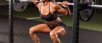
Wide Leg Squats
Exercises are aimed at strengthening the adductors, gluteal and quadriceps muscles:
- Stand straight, place your feet wider than your hips.
- Slowly lower your hips down as far as possible.
- Pause, tensing your internal muscles.
- Return to starting position.
- Do 2-3 sets of 8-12 repetitions.
Ball exercise
The gymnastic ball is suitable for strengthening the adductor muscles:
- Lie on the floor, knees bent, with a ball sandwiched between them.
- Squeeze the ball, alternately tensing your muscles.
Step sideways on one foot with the ball
To maintain balance, you can lean against a wall:
- Stand with your spine straight and place your hands on your sides.
- Extend one leg and place it on the fitball.
- While rolling the ball, slowly move your leg to the side.
- Repeat the exercise with the other leg.
Squat with dumbbells
Weighting exercise is aimed at strengthening the adductor muscles:
- Stand with your feet apart.
- Take dumbbells, lower your arms along your body.
- As you inhale, looking forward, squat down.
- When your hips are in a horizontal position, return to the original position.
- Exhale when finished.
Lunge with dumbbells
- Spread your legs slightly, take dumbbells, lower your arms, straighten your back.
- As you inhale, lunge wide forward with your left leg, place your right leg on your knee, and keep your torso straight.
- At the end of the movement, exhale.
- Repeat everything with the other leg.
Exercise "butterfly"
Movements help stretch the adductor muscles:
- Sit on the floor, back straight, bend your knees, connect your feet, heel to heel, toe to toe.
- Grab your feet with your hands, gradually moving them closer to the pelvis.
- Bend your back slightly forward and hold in this position.
- Try to lower your knees as much as possible.
- Hold the position for at least 3 minutes.
- During the exercise, breathe evenly and deeply.
- For every 4th exhalation, try to press your knees even closer to the floor.
- Slowly bring your thighs together and relax.
Leg scissors
This exercise helps stretch the inner thigh muscles:
- Lie on your back, raise your legs and bring them together. Extend your arms, place your palms down.
- Raise your legs and cross them in the air.
- Spread your legs all the way.
- Repeat the exercise, alternating the position of crossing your legs: first the right one in front, then the left one.
Seashell on its side
- Lie on your left side, stretch your body.
- Bend your knees, bring your hips closer to your body, not reaching 80-90˚. Place your left hand on your elbow, resting your palm on your head. The right one should be placed forward, the palm should be in front of the stomach.
- Lift the feet connected together from the floor, while the left thigh does not come off the floor.
- In this pose, the legs are spread and brought together at the knees.
- After 5-10 repetitions, turn to the other side and perform the exercise.
Read also[edit | edit code]
- Muscles - anatomy and functions
- Leg muscles
- Hamstring muscles
- Legs - exercises and training features
Muscles around the hip joint
- Gluteus maximus muscle
- Sartorius
- Iliopsoas muscle
- Gluteus medius muscle
- Gluteus minimus
- Tensor fascia lata
- Pectineus muscle
- Adductor longus muscle
- Adductor brevis muscle
- Adductor magnus muscle
- Gracilis muscle
- Superior gemellus
- Gemini inferior muscle
- Obturator internus muscle
- Obturator externus muscle
- Quadratus femoris
- Piriformis muscle
Recommendations
This article incorporates public domain text from page 473 of the 20th edition
Gray's Anatomy
(1918)
- Wilson, Erasmus (1851) Vade mecum anatomist: a system of human anatomy
, p.261 - MedicalMnemonic.com
: 255 - "Adductor Magnus." Department of Radiology, University of Washington. Archived from the original on August 28, 2010. Retrieved 2010-08-16.
- ^ a b
Platzer, Werner (2004),
"Color Atlas of Human Anatomy, Vol.
1, musculoskeletal system , Thieme, 5th ed., pp. 242. - Bergman, Ronald A.; Afifi, Adele K.; Miyauchi, Ryosuke (2010), Adductor Minimus (Henle, Gunther)
, Anatomical Atlases - Norman Eisenberg et al., Gross Anatomy: Principles and Applications
(2008), pp.
Anatomy, biomechanics, asanas for legs Part 2. Detailed analysis of the muscles of the lower limb
Abductor hallucis muscle (lat.Musculus abductor hallucis)
Description:
Located superficially, occupies the most medial position of the muscles of the plantar part of the foot.
It begins with two heads from the retinaculum of the flexor muscles (retinaculum mm.flexorum), the medial process of the tubercle of the calcaneus and the plantar surface of the scaphoid bone. Moving forward, the muscle passes into a tendon, which fuses with the tendon of the short flexor of the big toe (m.flexor hallucis brevis) and attaches to the medial sesamoid bone of the big toe at the base of its proximal phalanx Function:
flexes and abducts the big toe.
Participate in strengthening the medial part of the arch of the foot. Attachment:
origin - bones of the foot (medial process of the tubercle of the calcaneus and plantar surface of the navicular bone); attachment - to the base of the proximal phalanx of the big toe.
Flexor digitorum brevis (lat. Musculus flexor digitorum brevis)
Description:
muscle of the plantar part of the foot.
Occupies a mid-position on the foot, located under the plantar aponeurosis. It begins with a short powerful tendon from the medial process of the tubercle of the calcaneus and the plantar aponeurosis. Moving forward, the muscle belly passes into four tendons that lie in the synovial sheaths along with the tendons of the long flexor digitorum longus (m.flexor digitorum longus). In the area of the proximal phalanges of the II-V toes, each of the four short flexor tendons is divided into two legs, which are attached to the base of the middle phalanges of these toes. The tendons of the flexor digitorum longus pass between the legs. Function:
The muscle strengthens the arch of the foot and flexes the middle phalanges of the II-V toes.
Attachment:
origin - medial process of the calcaneal tuberosity and plantar aponeurosis; attachment - bones of the toes (bases of the middle phalanges of the II - V fingers)
Abductor muscle of the little toe (lat.Musculus abductor digiti minimi)
Description:
Located directly under the plantar aponeurosis.
It starts from the lateral and medial processes of the tubercle of the calcaneus and from the plantar aponeurosis. It goes forward and passes into a short tendon, which is attached to the lateral side of the base of the proximal phalanx of the little finger. Function:
Flexes the main phalanx of the little toe and pulls it laterally (abducts).
But the effect on the little finger is insignificant. The main role of the muscle is to strengthen the lateral edge of the arch of the foot. Attachment:
origin - bones of the foot (lateral and medial processes of the calcaneal tuberosity and plantar aponeurosis); attachment - the lateral side of the base of the proximal phalanx of the little toe.
Vermiform muscles of the foot (lat.Musculi lumbricales)
Description:
Four thin and short muscles that are located between the tendons of the long flexor of the fingers (m.flexor digitorum longus) and covered by the short flexor of the fingers (m.flexor digitorum brevis), and in depth they come into contact with the interossei muscles (mm.interossei plantares).
Each lumbrical muscle begins from the corresponding tendon of the flexor digitorum longus, with the three lateral ones (II-IV) having two heads, and the medial one (I) having one head. Moving forward, the muscles in the area of the metatarsophalangeal joints bend around the medial surface of the II-V toes and, moving to their dorsal surface, are woven into their dorsal fascia. Sometimes the lumbrical muscles are attached to the articular capsules and even reach the proximal phalanges. Function:
Flex the proximal phalanges of the II-V toes.
They have a weak or completely absent extensor effect on other phalanges of the same fingers. Can pull four fingers towards the thumb. Attachment:
origin - corresponding tendons of the long flexor of the fingers (m.flexor digitorum longus); attachment - bones of the toes (dorsal surface of the II-V toes).
Quadratus plantae muscle (lat.Musculus quadratus plantae)
Description:
The shape resembles a quadrangle and lies under the short flexor of the fingers (m.flexor digitorum brevis).
It starts from the lower and medial surfaces of the back of the calcaneus with two separate heads that connect into a common belly. Moving forward, the muscle narrows slightly and attaches to the outer edge of the long flexor tendon (m.flexor digitorum longus) at the site of its division into individual tendons. Function:
This muscle is like an additional head of the long flexor of the fingers (m.flexor digitorum longus).
This muscle bundle sets the longitudinal (straight) direction of traction of the flexor digitorum longus, the tendon bundles of which approach the fingers obliquely. In addition, the quadratus plantaris muscle increases the pulling power of the flexor digitorum longus muscle. Attachment:
origin - the lower and medial surfaces of the posterior part of the calcaneus; attachment - outer edge of the flexor digitorum longus tendon.
