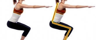
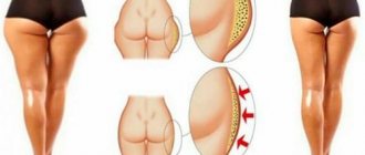
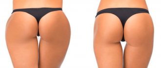
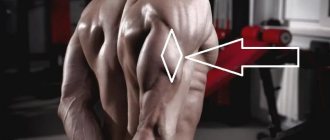
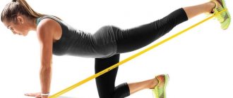
A common problem area for girls is the thighs, but sometimes large calves spoil the appearance. This may be due to swelling due to varicose veins, anatomical features, genetic predisposition, over-pumped muscles, as well as fatty layers in the lower leg area. Everyone dreams of graceful legs, and you can bring them to perfection with hard work. Special exercises will help girls reduce calves on their legs. Classes should be carried out systematically, following all the recommendations of specialists, temporary schedule and other rules.
- Seated stretching
Expert advice
When training at home, you need to remember the following nuances:
- Exercise regularly, systematically.
- Spend more time stretching your legs. Not only special exercises are suitable for this, but also gymnastics, Pilates, and yoga.
- You can do step aerobics: this will help fight the fatness of your calves. The lesson must last at least 40 minutes.
- It's good to run. You can use a special exercise machine or exercise outside.
- Sports walking and cycling will be beneficial.
How to eat healthy
Let’s admit right away: there is no separate diet for losing weight in calves. And this is natural. The body is our whole, and nutrition, be it right or wrong, will affect all parts of our body. A strict diet will only worsen the condition. Therefore, in pursuit of a slim figure, you need to eat right and healthy.
How many calories can you burn dancing?
First of all, the diet should be balanced
.
We include carbohydrates, fats and proteins. Of course, be sure to add vegetables and fruits
, which will fill the body with essential vitamins and fiber.
Carbohydrates
can be in the form of porridges, and if you can’t do without bread (do you already know about gluten?), then eat whole grain or yeast-free baked goods. Carbohydrates provide energy to our body and fight hunger.
Proteins –
a necessary building material for muscles (we have an article on this topic). They can be of both plant and animal origin. The main thing is to eat lean meat - baked, stewed or boiled.
The same goes for vegetables.
.
Not all vegetables can be consumed raw, but there are many wonderful recipes that will diversify your menu and make food consumption a daily holiday. Fresh salads are best seasoned with vegetable oil. It’s a matter of taste: olive, sunflower, flax and sesame are suitable. It’s not bad to add pumpkin, sunflower or sesame seeds to the salad. There are many meal options on sale now. We also recommend adding them to salads or cereals, since they are real weight loss products .
Don't forget about fresh fruit
.
Of course, it is better to eat fruits in accordance with the season
. Then they are maximally filled with vitamins and will bring you the most benefit.
We recommend following the basic principles of proper nutrition:
- fractional meals (often in small portions);
- healthy foods;
- sufficient amount of liquid (read more about how much water you need to drink);
- last meal no later than 3 hours before bedtime.
It should be noted that very often a large volume of calves can be a signal of swelling. To prevent water from retaining in the body, try to limit yourself to salty foods. And also eat foods containing potassium, as it absorbs sodium, which leads to swelling. Bananas, spinach, avocados, mushrooms, and chicken contain potassium.
Anatomical structure of the lower leg
Ankle structure
The leg consists of several complex structures:
- Femur. One of the strongest in the human body. It is powerful enough to withstand a load of more than 450 kg.
- The tibia and fibula are connected by thin, ribbon-like ligaments, which are very easily damaged when the ankle or other part of the lower leg is fractured.
- Calf muscles. They are not very pliable for forming relief. This is due to their constant work while walking and relatively little rest to build volume compared to the biceps of the arm.
There are a lot of ligaments in the lower limb that ensure the functioning of the foot and help maintain the vertical axis of the body.
The lower leg and foot are the most complex elements of the skeleton; they contain many ligaments that, intertwined, allow you to walk at the right pace and move smoothly.
Muscles and ligaments of the leg
The tibia consists of two bone formations:
- The tibia, which is located on the lateral part of the lower leg. Its diameter is on average twice the diameter of the fibula and it appears more massive. Most of the pressure from the femur is placed on the tibia.
- The fibula is located internally relative to the tibia and serves as a support for the vertical axis of a person. It is thinner than the fibula and therefore breaks more easily.
Calf muscles:
The back surface of the lower leg consists of the triceps muscle, which, in turn, consists of two smaller ones: the gastrocnemius and the soleus. The gastrocnemius muscle is represented by two heads: lateral and medial, and the soleus muscle is represented by one head.
The anterior surface of the lower leg is represented by several muscles: tibialis anterior, extensor digitorum longus and peroneus brevis.
The muscles of the anterior surface of the lower leg are quite thin, but their participation in the total volume of the lower leg is invaluable.
Recommendations for reducing calves on legs
Some girls got thick calves from their parents. In this case, the structure of the leg muscles is determined genetically, so it is very difficult to change it. But if the volume of the calves was initially small in childhood, and only increased with age, then all is not lost. Let's look at the reasons for voluminous calves and recommendations for solving the problem:
- Fat deposits. With a serious increase in body weight, the legs also become a problem area. Most of the fat is deposited on the hips, but the calves also get it. The solution is a low-carbohydrate diet and moderate exercise (aerobics, dancing, walking, mini-trampoline jumping, light fitness, Pilates).
- Excessive physical activity. Quite often, women involved in weightlifting, speed skating, and cycling have voluminous shins. To remove excess volume of calves on your legs, you will have to give up such activities. We suggest performing special exercises to lose weight in your calves (see below).
- Frequent wearing of uncomfortable shoes. Many women strive to look slimmer and sexier. To do this, they have to regularly wear high-heeled shoes. The result is regular stress on the calves and, as a result, excess volume. We recommend wearing comfortable shoes with low heels or flat soles more often.
- Edema of the lower extremities. The causes of the pathology may be heart failure, kidney disease, varicose veins, water-salt imbalance, and inflammatory processes in the body. In this case, in order to reduce the volume of the calves, it is necessary to undergo examination by a specialist.
- "Clogged" muscles. Women who spend all day on their feet may feel tightness in their shins in the evening. This visual “defect” is temporary. But if this happens every day, then over time the calves may become fuller. To make your calves smaller, we recommend relaxing them by stretching.
A little anatomy
The posterior group of muscles of the lower leg is represented by:
- External and internal parts of the gastrocnemius muscle;
- Soleus muscle.
The gastrocnemius muscle (also called the triceps muscle) is located above the soleus muscle and is attached to the heel using the Achilles tendon. These muscles perform important functions: the back surface of the lower leg produces forward and backward movement of the foot , and the anterior group of lower leg muscles provides it with a stable position while walking. These muscles, working together, bend the foot when walking. The calf muscles flex and extend the ankle joint and rotate it.
TOP 10 best exercises for legs - the most complete review in RuNet!
The calf muscles receive the greatest load during jumping, as well as when raising toes using weights. The soleus muscle gets a load when the knee is bent, so squats are a good way to train it. The gastrocnemius muscle is located above the soleus muscle - it is they who create the volume and shape of beautiful calves.
Massage
A good way to combat congestion in the ankles , which cause visual fullness of the calf muscles, is a comprehensive massage. Of course, this method cannot be called the only way out of the current situation, since it is better to use it in combination with stretching and physical exercise. To notice a rapid reduction in your calves, contact a professional massage therapist and undergo full-fledged therapy. According to experts, 6-10 sessions are enough for the patient to feel lightness of gait and feel comfortable.
However, in order to undergo massage prevention it is not necessary to go to a professional salon. There are many massage options that you can easily perform yourself at home. For example, lymphatic drainage massage, which consists of the following: take 2 chairs, sit on one, and put your foot on the second. The direction of the massage is from the feet to the knee, using leisurely and smooth movements. Immediately before the procedure, the skin should be treated with massage oil.
Is surgery necessary?
Among aesthetic surgeons, ankle liposuction is considered a highly controversial procedure. Some experts argue that ankle liposuction is useless because the shape and thickness of the ankles is determined by the genetic structure of the bone tissue.
Moreover, in this anatomical area there is not a large amount of fat, but there is a very high risk of damage to soft tissues and blood vessels. However, in some situations, specialists still agree to perform liposuction. But first of all, a consultation with a competent surgeon is necessary, who, after examining the patient, will determine whether there is a need for the procedure.
Surgery for ankle fractures
With external transosseous osteosynthesis, traumatologists use a guide apparatus with thin metal wires passed into the ankle joint to compare and fix bone fragments. The skin is damaged only in the area where the needle is inserted. Immersion osteosynthesis, carried out through an incision in the skin and soft tissues, involves the use of metal structures of various shapes and purposes, with the help of which fragments of damaged bones are connected.
In intraosseous osteosynthesis, rods are used, in bone osteosynthesis, plates with screws are used, and in transosseous osteosynthesis, pins and screws are used. During an open access operation, the traumatologist examines the damaged area in detail and also gets the opportunity to use the most effective surgical techniques. The disadvantage of this technique is heavy blood loss, disruption of tissue integrity, and the risk of wound infection.
Surgical techniques for ankle fractures
For fractures of the lateral (outer) ankle, the surgical incision is made in the projection of the fibula - along the outer surface of the ankle joint. After removing blood clots and small bone fragments, the surgeon repositions the fragments and then secures them with a plate and special screws.
Surgical treatment of injuries to the medial ankle involves two stages. The first is an incision along the inner surface of the ankle joint, cleaning the cavity from small fragments and blood clots. The second is to restore the integrity of the injured bone, fixing the fragments with knitting needles and screws.
The technique of surgical treatment of a bimalleolar fracture is determined by the condition of the articular fork and deltoid ligament. If the fork has retained its anatomical position (there are no signs of bone divergence), osteosynthesis of the medial malleolus is performed, then the lateral one.
A fracture of two ankles, complicated by divergence of the fork, is the basis for emergency surgery. First, osteosynthesis of the medial malleolus is performed, then a second incision is made along the fibula, after which osteosynthesis of the tibia is performed. The final stage of the operation is the application of a plaster cast.
A fracture of the anterior lower edge of the tibia with medial subluxation of the foot is a common injury in athletes. The surgical technique is as follows: a long longitudinal incision is made, cutting the transverse and (sometimes) cruciate ligament, the tendons are pulled apart with blunt surgical hooks to expose the site of bone damage. The foot is bent and shifted back, the fragments are set, connecting them with metal rods (the pin is driven into the tibia). Next, the foot is extended and placed at a right angle. The hooks are removed, tissue is sutured layer by layer, and a plaster cast is applied to the knee.
A fracture of the lower posterior edge of the tibia with posterior dislocation of the foot is a difficult case in traumatology. The operation is urgent. The patient's position is face down. The incision is strictly parallel to the Achilles tendon, along the outer edge. After exposing the injured area, the fragments of the tibia are set, fastening the joining area with a screw or a special nail. The reduced foot is brought into a vertical position (at a right angle to the lower leg). With this type of fracture, it is technically difficult to remove metal structures after restoration of the joint, therefore, if possible, the technique of external transosseous osteosynthesis is used.
Metal fixators are removed 3-6 months after osteosynthesis. A full-fledged surgical operation is performed.

