The buttocks, as the gluteus maximus muscle is called (musculus gluteus maximus), many of us rarely attach importance to, and do not consider these muscles important, and remember them only when the swimming season comes, and the need arises to wear swimming trunks and, moreover, attention is paid form, not function. The gluteal muscles serve several functions beyond just being an attractive seat cushion, and if these muscles are weakened, then a lot of problems arise. Well, if we sit all the time during the day (at work, at home, while eating or watching TV), then what kind of muscle strength can we talk about?
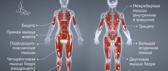
Nature made this muscle large enough not only for the sake of beauty. The gluteus maximus muscle (“MGM”) is the largest and one of the strongest muscles in the human body. The gluteus maximus muscle is the most superficial of the three gluteal muscles and forms most of the shape and appearance of the buttocks. The gluteus maximus muscle runs along the crest of the pelvic bone and attaches to the posterior aspect of the proximal femur and to the iliotibial ligament, providing a connection between the trunk and lower extremities.
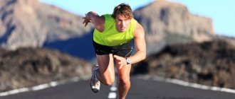
The large size of this muscle is one of the most characteristic features of the human muscular system. According to most researchers, humans evolved from ape-like ancestors about two million years ago on the vast savannah of Africa. It is believed that natural selection favored the survival of animals that could escape. Over time, evolution consolidated the anatomical features that allowed humans to run long distances, and the enlargement of the gluteus maximus muscle may have played a decisive role in this process.
Functions of the gluteus maximus muscle.
The gluteus maximus muscle is very important in performing activities such as standing, walking, and running. The function of the muscle in these activities is to participate in straightening the leg, bringing the torso into a vertical position, abducting and adducting the hip in accordance with our body, rotating the hip from and to the center of the body and stabilizing the pelvis. This muscle may also play a role in stabilizing the knee during extension.
During standing up, for example, the gluteal muscle plays an important role in hip extension and pelvic stabilization. During running, this muscle provides stabilization to the torso and helps extend the hip when accelerating and slow down the leg when stopping.
Thus, weak gluteus maximus muscles reduce the ability to perform many of our daily activities effectively and safely. Muscle weakness can make it difficult to perform certain movements that require these muscles, such as standing up, sitting, walking, or running. Sometimes weakness of the gluteal muscles can be associated with back, knee and hip pain.
Read also[edit | edit code]
- Muscles - anatomy and functions
- Leg muscles
- Hamstring muscles
- Legs - exercises and training features
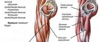
Muscles around the hip joint
- Gluteus maximus muscle
- Sartorius
- Iliopsoas muscle
- Gluteus medius muscle
- Tensor fascia lata
- Pectineus muscle
- Adductor muscles
- Adductor longus muscle
- Adductor brevis muscle
- Adductor magnus muscle
- Gracilis muscle
- Superior gemellus
- Gemini inferior muscle
- Obturator internus muscle
- Obturator externus muscle
- Quadratus femoris
- Piriformis muscle
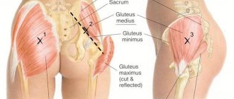
How do you know if your gluteus maximus muscles are weak?
For example, here is a simple and quick test that will help determine if you have weak gluteal muscles.
Lie on your back with your knees bent at least 90 degrees. Squeeze your gluteal muscles. Slowly lift your hip off the floor and then straighten your right knee so that your thighs are parallel. Try not to change the level of your pelvis or rotate your hips. If the pelvis deviates or rotates to one side when the leg is extended, then there is definitely weakness in the gluteal muscle. But this test should not be performed by women until at least 6 weeks after giving birth.
Exercise therapy doctors can use additional manual and dynamic diagnostic methods to determine gluteal muscle weakness:
- Manual diagnostic techniques include observing whether the exercises are performed correctly, whether there are any displacements in the movement, or whether other muscles are compensatory in the movement.
- Dynamic methods include digital analysis of video of movement performance, which allows the exercise therapist to examine the movement in detail. Such video analysis allows the physical therapy doctor to evaluate the work of the gluteal muscles in the kinetic chain, to observe the function of the pelvis and lower extremities dynamically, throughout the entire gait cycle.
Pathology
Pathologies of the gluteal region include injuries, inflammatory diseases, and tumors. A rare type of pathology of the gluteal region is a sciatic hernia.
Damage
There are closed and open injuries to the gluteal region. Closed injuries - bruises (see) occur when struck or fallen. In this case, damage to the subcutaneous tissue, muscles and blood vessels occurs, the nature and extent of which depend on the strength of the impact and the area of damage. As a rule, bruises are accompanied by the formation of extensive subcutaneous, subfascial and intermuscular hematomas (see), sometimes causing detachment of the skin or subcutaneous tissue. With prolonged crushing of the soft tissues of the gluteal region, especially in combination with crushing of the tissues of the lower extremities, traumatic toxicosis can develop (see). If the gluteal arteries are damaged during a bruise, extensive interstitial hemorrhages occur, spreading through the interfascial spaces into the pelvic cavity and onto the thigh, up to the popliteal fossa. Such hemorrhages can lead to significant blood loss. Sometimes the hematoma and blood-soaked tissue can encyst to form a traumatic cyst filled with hemolyzed blood (see Hematoma). When calcium salts are deposited in damaged and blood-soaked muscles, traumatic myositis ossificans develops, which persistently impairs muscle function (see Myositis). Severe dysfunction of the muscles of the gluteal region in traumatic myositis ossificans is an indication for surgical intervention - excision of ossified tissue.
Injuries to the gluteal arteries and open injuries to the gluteal region most often occur with combat gunshot wounds. In peacetime, lacerations and stab wounds occur as a result of a fall or injury. In this case, sometimes there is a bruise, partial or complete rupture of the sciatic nerve (see), which is clinically manifested by paresis and paralysis. Particularly difficult are combined injuries of the gluteal region and pelvic organs, as well as wounds penetrating through the tissues of the gluteal region into the pelvic cavity. In these cases, injury to the bladder is possible, accompanied by urinary leakage into the gluteal region (see Urinary leakage), as well as injury to the rectum (see), leading to the rapid development of purulent putrefactive phlegmon of the pelvis and gluteal region (see Pelvis, injuries ).
Combat injuries to the gluteal region include gunshot wounds, which can be tangential, blind or penetrating. Blind wounds in this area, both bullet and shrapnel, are more common than with wounds in other areas. With gunshot wounds of the gluteal region, damage to the superior and inferior gluteal arteries is possible, accompanied by heavy external bleeding. With a long and narrow wound channel, bleeding from pinpoint wound openings is insignificant, and therefore vascular damage may go unnoticed. In these cases, as with subcutaneous ruptures of the arteries, conditions are created for the formation of bursting or pulsating hematomas. Diagnosis of injuries to the gluteal arteries is difficult. Initially, due to the blood soaking of surrounding tissues, a diffuse infiltrate is formed. Usually, the pulsation of the hematoma can be detected only 5-12 days after the injury, since the source of bleeding is covered by a massive layer of the gluteal muscles. When a hematoma compresses the sciatic nerve, pain appears, which increases with the development of a wound infection. In these cases, a pulsating hematoma is often mistaken for a purulent infiltrate. With blind wounds of the gluteal region, a large fragment can put pressure on the sciatic nerve, which sometimes leads to the development of painful shock (see). According to the experience of the Great Patriotic War, gunshot wounds of the gluteal arteries accounted for 0.4-4% of all injuries to blood vessels. The superior gluteal artery was most often damaged (67.8%). In 76.1% of the wounded, damage to the gluteal arteries caused secondary bleeding; in 23.9%, pulsating hematomas and aneurysms were observed. Gunshot wounds of the sciatic nerve ranked second in frequency after injuries to the radial nerve and accounted for 14.9% of all nerve trunk injuries. Damage to the sciatic nerve is sometimes combined with injuries to the gluteal arteries. Isolated injuries to the sciatic nerve in themselves are not life-threatening, but sharply reduce the likelihood of the wounded person returning to duty.
Gunshot wounds and open injuries to the gluteal region, in addition to the possibility of damage to its arteries and nerve trunks, pose a great danger due to the development of infectious complications, especially anaerobic infection (see). A large amount of fatty tissue and muscle tissue, as well as their relatively poor blood supply, cause a severe course of anaerobic infection and purulent complications, which are accompanied by the formation of numerous streaks and secondary putrefactive infection. This often leads to the development of sepsis (see).
Treatment of closed injuries of the gluteal region is conservative: bed rest, painkillers; for extensive hemorrhages, antibiotics are prescribed for prophylactic purposes. In the first 2-3 days, cold is applied topically, followed by physiotherapy (UHF, diathermy). In case of traumatic detachment of the skin or subcutaneous tissue, apply a pressure bandage and remove the accumulated fluid under the skin using a puncture. If the puncture is unsuccessful, a skin incision is made and the fluid is removed. Non-viable skin and subcutaneous tissue are excised, followed by replacement of the defect with a free skin graft. In case of closed damage to the artery, characterized by a rapidly growing hematoma, deterioration of the general condition, and a decrease in the hemoglobin content in the blood, an operation is performed to stop the bleeding. In case of open damage, surgical treatment of the wound is performed (see), and the damaged sciatic nerve is sutured (see Nervous suture, Sciatic nerve).
When surgically treating a wound, stopping bleeding presents significant difficulties. Ligation of the gluteal arteries in the wound to completely stop bleeding is technically difficult and is associated with additional trauma. It is not possible to grab the short trunk of the arteries that still branch in the pelvis with a hemostatic clamp, even after a wide incision along the fibers of the gluteus maximus muscle. Therefore, if it is necessary to stop bleeding, it is more advisable to start it with extraperitoneal ligation of the internal iliac artery. The operation is performed under general anesthesia. Before ligating the internal iliac artery, the wound in the gluteal region is carefully packed. The Shevkunenko operative approach is used: an arcuate incision is made from the end of the 11th rib downwards and anteriorly to the superior anterior iliac spine, and from the latter - medially. The peritoneum is peeled off bluntly; the internal iliac artery, located 1 cm medially from the external iliac artery, is ligated with two silk ligatures. Collateral circulation is restored through anastomoses of the branches of the femoral artery with the superior and inferior gluteal arteries and with the obturator artery. Ligation throughout, as a rule, ensures the final stop of bleeding from the gluteal arteries. Even if bleeding continues from the peripheral end of the gluteal artery due to the opening of collateral blood flow, it is reduced so much by ligation that the search for a bleeding vessel in the gluteal region can be continued without the risk of losing the patient on the operating table from ongoing bleeding. In this case, to finally stop the bleeding, the incision proposed by A.G. Radzievsky for approaching the damaged sciatic nerve is more convenient, in which the gluteus maximus muscle is crossed along the outer edge and turned inwards. At the end of the operation, the gluteal muscle is sutured, starting from the bottom. The most common complication in the postoperative period is wound suppuration.
The principles of staged treatment of wounded with injuries to the gluteal region are the same as when providing assistance to wounded with injuries of the pelvis and pelvic organs (see Pelvis). First aid consists of applying an aseptic pressure bandage to the wound and administering painkillers. When providing first aid (see), the bandages are monitored and corrected, and painkillers are administered. The victim is transported on a stretcher. First medical aid consists of taking measures to prevent shock (see) and control bleeding (see). Bleeding is stopped by tight tamponade of the wound (see Tamponade), and it is possible to apply temporary sutures to the skin over a tampon, which must be removed no later than 24 hours, and even earlier if signs of infection appear. When providing qualified surgical care after medical triage (see), all wounded in the gluteal region are divided into the following groups: wounded with ongoing bleeding, subject to urgent surgical intervention; wounded in a state of shock without signs of bleeding, who are receiving anti-shock therapy; wounded in soft tissues that do not require emergency surgical intervention, who undergo primary surgical treatment of the wound and operations on blood vessels in case of damage. Specialized care includes surgical interventions for developed infectious complications, traumatic aneurysms of the gluteal arteries and injuries to the sciatic nerve. It is often necessary to operate due to late bleeding from the gluteal artery, damage to which often remains unrecognized at the previous stages of evacuation.
The main method of treating anaerobic infection of the gluteal region is surgery, which consists of a wide dissection of the wound, opening of the muscle fascial sheaths, excision of necrotic tissue, removal of foreign bodies and loose bone fragments, opening of blind pockets and depressions running away from the wound, leaving the wound wide open. When an anaerobic infection occurs, in addition to surgical intervention, the administration of anti-gangrenous serum and antibacterial agents, timely oxybarotherapy is of great importance, which can significantly improve treatment results (see Hyperbaric oxygenation).
What can you do to increase glute muscle strength?
There are many exercises you can do that will not only improve the shape and tone of your glutes, but also improve their function.
One leg bridge
Lie on your back with your knees bent at least 90 degrees. Squeeze your gluteal muscles. Slowly lift your hip off the ground and then straighten your right knee so that your thighs are parallel. Keep your pelvis level and don't rotate your hips. Hold for 10 seconds and then return to the starting position. Repeat this exercise on your left leg. Repeat the exercise 10 times. Note: This exercise should not be performed by women until at least 6 weeks after giving birth.
Lying hip extension
Lie on your stomach, 1-2 pillows under your hips. Bend your knee 90 degrees. Squeeze your glutes and slowly lift your heel toward the ceiling. Don't let your back sag. Hold your leg in this position for 10 seconds and then return to the starting position. Repeat 10 times. Note: if you have back pain, then this exercise can only be performed under the supervision of a physical therapy doctor.
Clinical relevance
Weakness of the MMN leads to the development of a Trendelenburg gait (the pelvis drops on the side opposite to the supporting leg when walking).
MMN tendinopathy is associated with greater trochanter pain syndrome, which is characterized by pain radiating down the lateral thigh, tenderness of the greater trochanter, and a Trendelenburg gait. This condition should be differentiated from greater trochanteric bursitis.
PMN trigger points can cause referred pain that begins in the lumbar region, travels down the leg and ends in the calf. It is often similar to the pain of sciatic nerve irritation, but without neurological symptoms such as weakness and numbness.
Bret Contreras
Strength Coach and Performance Expert
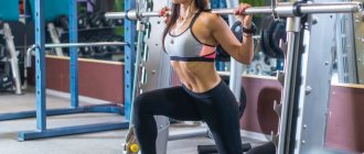
To pump up the gluteal muscles, you need to meet two conditions: load them quite heavily (and increase the load over time) and learn to mentally concentrate on the working muscle.
Start your workout with a “heavy” exercise that allows you to move heavy weights—such as a barbell squat, Romanian deadlift, sumo deadlift, deadlift, or platform leg press. Keep a training diary and regularly record your progress in these exercises - weights, sets, repetitions. It is important to get enough rest between sets.
Finish the workout with lighter, isolation exercises such as frog pumps with dumbbells, glute bridges with a resistance band around the knees and dumbbells, hyperextensions without weights and the like.
Do more repetitions of these exercises and rest less in between. You don't have to count reps, but it is important to engage your glutes as you work and focus on the quality of each rep.
The combination of these two approaches allows you to work on your glutes with maximum impact.
Mark Dugdale
IFBB Pro Bodybuilder
My best advice for anyone who wants strong, great-looking glutes is to start every leg workout by activating and pre-exhausting your glutes. You'll see they perform better throughout the rest of your workout.
Here are a few of my top suggestions for butts:
- Glute Bridge: Pause each rep for 2 seconds at peak muscle contraction.
- Split squats with one leg on the bench: do drop sets (reducing the working weight several times per set as the muscle fatigues) with a pause of a few seconds at the end of the set.
- Prowler push: Take long, slow, deliberate strides using moderate weights on a sled.
Try starting every leg workout with one of these exercises. Alternate between them because they all activate the glutes differently.
Causes
Myth #1 Quote from a reputable resource: “Many people believe that this (the occurrence of piriformis syndrome) occurs when the piriformis muscle goes into spasm and begins to press the nerve against the pelvic bone.” - Yes, this is definitely the reason for the pain, but the reason for the syndrome is that it caused painful contraction and inflammation in the piriformis muscle .
There are quite a few reasons for this unpleasant condition:
- Primary disruption of the piriformis muscle (eg, failure of an injection to puncture the piriformis muscle)
- Inflammatory phenomena in the hip joint (for example, with coxarthrosis)
- Diseases of the pelvic organs (genitourinary system)
- Problems in the sacroiliac joint and sacrum (injuries, neoplasms)
- Incarceration of the spinal roots of the lumbar and sacral spine
- True inflammation of the sciatic nerve (quite rare)
Contrary to popular belief, external factors do not have a strong influence on the piriformis muscle : it is protected from shock and low temperatures by the gluteus maximus muscle.
Bronwen Blunt
Nutrition and Strength Coach
You can't build a house without a foundation, so start with the "big" compound exercises - squats, deadlifts.
I see a lot of bikini athletes doing endless glute isolation and completely neglecting compound exercises in their programs. You won't build big, round buttocks by sitting on machines and doing endless reps all day. Build the foundation first, and after that, all other exercises will be more beneficial for your figure.
The three main exercises for strengthening and growing the buttocks are barbell squats, sumo deadlifts, and Bulgarian split squats.
Try adding some manipulation to your exercises to further challenge your muscles. For example, hold the glute contraction for 3-5 seconds at the top of the sumo lift at the end of each rep, or increase the negative phase (lowering the weight) to 5 seconds.
Diagnosis of piriformis syndrome
To recognize piriformis syndrome, methods such as:
- Certain manual tests
- Neurological tests
- Infiltration of the piriformis muscle with novocaine with assessment of the resulting positive changes
Recognizing piriformis syndrome is not difficult, it is much more difficult, but it is absolutely necessary to find out its true cause. The idea that piriformis syndrome may be the final diagnosis is another myth.
Examination of the piriformis muscle itself can be difficult, so if there is no indication of damage to it, it is examined last.
If piriformis syndrome is confirmed, the study begins with an MRI of the lumbar spine and ultrasound of the pelvic organs. An X-ray examination of the hip joint can then be performed to possibly detect coxarthrosis at an early stage.