What a beginner needs to know before starting to lose weight
Before you start losing weight, a beginner should know that it is impossible to transfer fat into muscle. This means that if a person is overweight, then when he starts working out in the gym, he will not achieve the effect of muscle accumulation and fat breakdown.
In other words, in order to remove fat tissue, you need to use some processes, and in order to build muscle, others.
If the goal is to lose weight and get pumped up, you won’t be able to do it at the same time. It is impossible to drive one cell into another, as some people think. First you need to lose weight by losing excess weight, and only then build muscle.
Wraps, massages, rubbing will not help you lose fat mass. Fat from the triglycerides in which it is found in the body will be broken down into fatty acids and glycerol will enter the bloodstream. But if you don’t use it, for example, don’t give physical activity or don’t reduce calorie intake, these cells will return to their original place, accumulating on the sides. They can also deposit plaques on blood vessels in the form of cholesterol.
Eating right is very important
You can only get rid of excess fat if you eat right. This is exactly what a beginner who has decided to say goodbye to extra pounds should know. To do this, you can play sports, run, jump, swim, that is, do cardio exercises, but only slowly and for more than an hour at a time. You should train every other day. This will be enough, but if problems with obesity are serious, then it is better to exercise every day.
Having lost the hated kilograms, you can move on to gaining muscle mass. Then, perhaps, the kilograms will return, but the person’s body will look many times more beautiful. It will become slim and fit
To lose fat and gain muscle, you need to eat differently. In the first case, the amount of carbohydrates should be reduced to a minimum or completely abandoned for some time. In the second case, you should eat a limited amount of carbohydrates in the first half of the day, and increase your protein intake in the second half of the day. The general thing is that you need to eat little by little and in fractions.
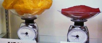
Important! Only by knowing what 1 kg of fat and 1 kg of muscle looks like can you build up the former and fight the latter.
Adipose tissue is present in varying quantities in all tissues of the body, and its distribution depends on many factors, including gender, age, ethnicity, genetic characteristics, level of physical activity, and diet. With the development of new high-resolution imaging technologies, it has become possible to assess both the total amount of adipose tissue in the body and study the topographic features of its distribution.
Currently, there are two subtypes of intra-abdominal adipose tissue with different metabolic characteristics: subcutaneous (SAT) and visceral (VAT). Of particular interest is the study of visceral deposition of fat mass, since it has been shown to be associated with a high risk of developing cardiovascular diseases, type 2 diabetes mellitus (T2DM), insulin resistance (IR) and dyslipidemia. BAT is a source of a number of biologically active peptides and adipokines (visfatin, etc.). Adiponectin is a unique “cardioprotective adipokine”, a decrease in the serum level of which with the progression of obesity is an independent risk factor and predictor of cardiovascular diseases, metabolic syndrome (MS) and T2DM [1, 2]. Serum adiponectin levels are inversely proportional to the amount of BAT and negatively correlate with the main components of MetS [3]. Thus, it is visceral obesity that determines high cardiometabolic risk, and the assessment of the total amount of adipose tissue and BAT (simultaneously and with dynamic observation) becomes particularly relevant.
There are many methods for determining the amount and distribution of fat tissue in the body. The most common in clinical practice are those that do not require much time and give quick results. These include anthropometric methods and bioimpedance analysis. Along with simple ones, there are more technologically advanced imaging techniques, such as computed tomography (CT) and magnetic resonance imaging (MRI), during which direct visualization of adipose tissue and its quantitative assessment are possible. However, these methods are labor-intensive and expensive, which limits their widespread use in clinical practice. Each method has its own advantages, disadvantages and scope of application, which will be discussed in this review.
Anthropometric methods
Anthropometric methods are the simplest and most accessible for assessing the amount of fat mass in the body.
Body Mass Index (BMI)
is the most widely used method in clinical practice that characterizes the presence of obesity and its severity. In adults, a BMI <25 kg/m2 indicates overweight, and a value <30 kg/m2 indicates obesity.
H. Kvist et al. [4] showed that BMI correlates with total adipose tissue, but not with BAT. The disadvantages of BMI are the inability to estimate the amount of lean and visceral fat mass. In children, the use of BMI is even more difficult, since it becomes necessary to make adjustments for gender and age (i.e., use percentile tables).
Waist circumference (WC)
is another anthropometric indicator widely used in the adult population to indirectly assess visceral obesity. There are at least 7 different methods for measuring WC. It is measured at the midpoint of the distance between the lower rib and the edge of the iliac crest according to the criteria of the International Obesity Group and WHO (IOTF, WHO, 1997).
WC has been shown to correlate with the amount of BAT determined using CT and MRI. According to the recommendations of the NCEP ATP III (National Cholesterol Education Program Adult Treatment Panel III), WC <88 cm in women and <102 cm in men is a sign of abdominal obesity and is associated with high risk of developing diseases. The International Diabetes Federation (IDF) has established a WC limit of <94 cm in men and <80 cm in women, respectively. Several countries have developed national percentile tables for assessing WC in children. It should be noted that WC values characterizing visceral obesity in the pediatric population are significantly lower than in adults [5].
OT/OB ratio and sagittal diameter
(abdominal height with the patient in the supine position) are additional indicators used in clinical practice to assess the distribution of adipose tissue (AT) in the body. It has been shown that WC and sagittal diameter reflect the degree of visceral obesity, while the WC/HR ratio (ratio of waist and hip circumference) reflects the degree of development of subcutaneous fatty tissue. The value of the OT/OB coefficient <1 in men and <0.85 in women indicates the predominance of VAT. J. Kullberg et al. [6] showed that BMI and sagittal diameter were positively correlated with the amount of BAT measured by MRI.
All anthropometric methods are indicators of VAT accumulation, but do not allow quantitative assessment of it. The advantages of all anthropometric methods include the ease of their implementation and wide availability. However, they all have large measurement errors. Therefore, more accurate methods are needed to estimate the amount of fat mass and BAT.
Bioimpedance analysis
Bioimpedance analysis (BIA) is an electrophysical method based on measuring the electrical resistance of tissues of the whole body and its individual parts. Based on the resistance value, the total water content in the body is initially calculated, and then, using mathematical algorithms, the amount of lean mass is calculated. The amount of fat mass is obtained by subtracting lean mass from total body mass. Thus, the quantification of VT using BIA will be influenced by the amount of body water, which depends on gender, age, level of physical activity and water load, as well as the error in measuring body weight. All these factors significantly reduce the accuracy of this method. When comparing the results obtained using BIA with data from dual-energy X-ray absorptiometry (DXA) in adults, good comparability of these methods in determining the amount of fat and lean mass is shown [7-10]. BIA somewhat overestimates the amount of adipose tissue in the body compared to DXA, but quite accurately reflects the content of lean mass, especially in the absence of obesity [11].
The advantages of the method are its low cost and availability, lack of radiation exposure and the ability to conduct studies over time. BIA allows you to assess both the total amount of fat and lean mass in the body and its regional distribution. Like anthropometric methods, BIA does not make it possible to directly assess the amount of BAT and PVT, however, the latest generations of body composition analyzers make it possible to mathematically calculate the volume of BAT. In terms of its ability to assess the total content of fat mass in the body, this method is significantly superior to anthropometric methods, but is significantly inferior to other high-resolution methods.
In pediatric practice, there is no uniform standard for conducting research and standards for various BIA indicators. Data on the comparability of the results of BIA and DXA in children are extremely contradictory. A number of authors [11-14] point out their comparability, especially when determining the amount of lean mass in both obese and non-overweight children. Others [15, 16] believe that these methods are not equivalent and not interchangeable, and for the pediatric population it is necessary to develop separate standards depending on gender, age and stage of sexual development.
In 2012, Kai-Yu Xiong et al. [17] conducted BIA in 1548 children and adolescents and for the first time determined the standards for the amount of fat and lean mass in the Chinese population depending on age and gender. Less than 1 year later, H. McCarthy et al. [18] created percentile curves to estimate lean mass and relative and absolute muscle mass for a European population using bioimpedance analysis in 1985 nonobese and overweight children and adolescents. Further conduct of such studies will make it possible to develop uniform standards for assessing body composition indicators using the BIA method depending on gender and age in the pediatric population.
Thus, BIA and anthropometric methods may be useful in clinical practice for the initial diagnosis of obesity and determination of the type of fat distribution.
Ultrasonography
Ultrasound examination (US) is widespread in various fields of medicine.
With the development of technology and improvement in resolution, attempts have been made to assess the amount of PVT and VAT using ultrasound. The method for determining the thickness of mesenteric fat consists of measuring the distance between the anterior abdominal wall (rectus abdominis muscles) and the anterior wall of the aorta at a level of 5 cm below the xiphoid process [19]. The thickness of VT at which VAT is diagnosed, according to various sources, ranges from 7 to 11 cm. Data on the comparability of ultrasound assessments of VAT and PVT with the results of reference methods are provided. Other authors [20] report low accuracy of measurements of PVT and VAT, which do not coincide with CT or MRI data. In addition, the disadvantages of ultrasound include the high variability of results during repeated studies, especially by different specialists.
Dual-energy X-ray absorptiometry
Dual-energy X-ray absorptiometry (DXA) is a radiation diagnostic method based on recording the attenuation of X-ray radiation when passing through body tissues of different densities. The device’s software calculates the attenuation of X-ray radiation and, using specified algorithms, determines the amount of tissue of one density or another. DXA is the gold standard for assessing bone mineral density and diagnosing osteoporosis. However, the whole body scanning module present in modern X-ray densitometers allows one to assess not only skeletal mineral density, but also the amount of fat and muscle tissue, as well as their regional distribution. The examination is carried out in the supine position; the duration of scanning the whole body ranges from 10 to 30 minutes, depending on the device used. The radiation exposure during the study is 10 times less than what the patient receives during standard radiography of the lungs and amounts to 0.02-0.04 mSv per scan. This method is easy to perform and does not require much time or special preparation of the patient. In addition, the minimum radiation dose allows the use of DXA when conducting dynamic studies. The accuracy of assessing the amount of adipose tissue is comparable to that of CT and MRI [21]. All these advantages lead to the widespread use of DXA in scientific research on the problem of obesity.
Currently, there is conflicting data on the correlation of DXA indicators with the amount of visceral fat. A. Hill et al. [22] showed that the area of VT, assessed using CT analysis of individual slices at the level of L4-L5, strongly correlates with the percentage of VT in the trunk, determined using DXA, but other authors [23] did not find similar relationships. This may be due to the fact that DXA differentiates between tissues based on density. VT has the minimum density, and bone has the maximum. When assessing a region of the body that contains predominantly bone and fat, the software averages the tissue density of the region of interest and classifies it as lean mass, which may influence the estimate of total fat mass. A number of works [24, 25] are devoted to the regional assessment of adipose tissue in the area 5-10 cm above the iliac crest, since this zone is traditionally used to assess visceral obesity using CT and MRI methods. However, such measurements have low accuracy and cannot currently be used as an alternative to CT and MRI. Despite certain shortcomings, DXA is a significantly more accurate method for assessing VT than anthropometry and BIA.
Computed tomography (CT) and magnetic resonance imaging (MRI)
Due to their high resolution, these methods allow direct assessment of the amount of BAT and PVT in adults and children [26]. CT is a method of radiation diagnostics based on the calculation of the attenuation coefficient of X-ray radiation intensity when passing through tissue, expressed in Housefield units. The imaging range of adipose tissue is from –190 to –30 Housefield units [27]. One of the challenges in assessing VAT using CT and MRI is the choice of slice level at which to determine the area occupied by visceral VT. G. Borkan et al. [28] proposed to determine VAT at the level of the navel, since the maximum number of VAT corresponds to this level. However, measurement at the level of the umbilicus is extremely inaccurate due to the variability of this anatomical landmark depending on the degree of obesity. Therefore, it was recommended to use bony landmarks to determine the level of cut. This reference point was the level of the L4-L5 intervertebral disc. VAT studies at this level show maximum reproducibility and high accuracy of measurements. A number of studies [29, 30] have shown that the assessment of VAT at 5-6 cm above L4-L5 is more accurate when performing dynamic MRI and CT.
Despite a large number of studies, there are currently no uniform criteria for assessing the presence and severity of visceral obesity. A number of authors [31, 32] indicate that visceral fat mass area <100 cm2 in both men and women and a BAT/PVT ratio <0.4 are associated with a significant increase in the risk of cardiovascular diseases. Other authors [33] suggest a figure of 130 cm2.
The main problem in using CT and MRI to evaluate VT is motion artifacts. In addition, CT is associated with radiation exposure, which limits the use of this method in the pediatric population, especially when long-term dynamic observations are necessary. The cost of the equipment and the test itself, as well as the training of specially trained personnel, also limit the use of CT for the assessment of VAT in routine clinical practice.
MRI is another high-tech modality for imaging intra-abdominal and ectopic VT in both adults and children. When studying VT, T1-weighted spin echo sequences are used, since VT, unlike most “non-fat” tissues, has a high T1-weighted signal and is well visualized in this mode. Quantitative assessments of BAT using MRI are comparable to CT data [26]. In MRI, in addition to T1-weighted images, other pulse sequences can be used: 3D gradient echo sequence and fast Dixon sequence, which reduce examination time and increase its information content. MRI is much more susceptible to motion artifacts associated with respiration and cardiac activity than CT. Their number can be minimized by fixing the part of the patient’s body being examined, synchronizing tomography with ECG and breathing, and using fast imaging (FISP - fast imaging with steady-state precision) [34].
Currently, it is possible to perform MRI with the acquisition of individual and multiple slices through certain anatomical levels, followed by multiplanar reconstruction and determination of the number of VT. An MRI scan of individual sections scans the body at the same levels as a CT scan. Next, the image is reconstructed using software. A radiologist experienced in assessing VT manually marks the VT and PVT and then automatically calculates the area occupied by each (see figure).
Picture 1.
Figure 3. MRI examination of intra-abdominal VT. a — original MR image (single section at the L4-L5 level); b — VAT; c — PVT.
Thus, the qualifications of a radiology specialist play an important role in assessing the amount of adipose tissue using this method. Another limiting condition for MRI is the difficulty in using these methods in patients with morbid obesity due to the load limit on the tomograph table (usually no more than 130 kg) and a large waist circumference, incompatible with the diameter of the scanner tunnel. Multi-slice MRI allows assessment of the volume occupied by intra-abdominal VT. In this case, the body is scanned from level Th 9-10 to S1. Subsequently, each of the slices is evaluated separately and the data obtained are summarized, which makes it possible to calculate the volume of VT, and not just its area, as in an MRI study of one slice. This method requires significantly more time, which significantly limits its widespread use. In addition, the analysis of individual MRI sections is comparable in accuracy to the assessment of VAT with the calculation using the method of multiple MRI sections in a simultaneous study. However, their accuracy is insufficient to assess the dynamics of the amount of BAT during long-term observation during weight loss. This circumstance dictates the need to create fully automated software for assessing the amount and topography of VT using MRI. Such algorithms will ensure a reduction in the time spent on MRI studies with evaluation of multiple slices and will significantly reduce its cost, which will help increase the availability of this technique.
Conclusion
Currently, there are many methods to assess the amount and distribution of VT in the body.
Anthropometric methods are the simplest, most convenient and cheapest, and therefore are widely used in clinical practice. Their main disadvantages are the inability to assess the amount of lean mass and differentiate between BAT and PVT. OT can be used to indirectly assess BAT in adults, but the informative value of this method in pediatric practice remains controversial.
BIA allows you to assess not only the amount of lean and fat mass, but also their regional distribution. The accessibility and simplicity of the method make it possible to use it for dynamic monitoring of obese patients and assessing the effectiveness of ongoing treatment measures.
The main disadvantages of the method include low reproducibility of results and the inability to estimate the amount of VAT.
Ultrasound allows direct visualization of VAT, but there is no convincing data on the comparability of ultrasound results with data from reference methods. In addition, ultrasound does not assess the total amount of fat and lean mass in the body. X-ray densitometry in Total Body mode is the “gold standard” for assessing the amount of fat and lean body mass and is used in most clinical studies on obesity. The comparability of quantitative assessments of VAT using this method with the results of reference methods (MRI and CT) has not currently been studied.
MRI and CT are the reference methods for assessing the amount of VAT and PVT. However, the high cost and radiation exposure of CT scans limit their use to research. MRI is a more promising method for assessing visceral obesity in clinical practice, especially in children.
Human Fat Density and Muscle Density
Abdominal muscles
Muscle has a higher density than fat. It turns out that greater density occupies a smaller volume of the same weight than less density, and a person who has a lot of muscle tissue looks slimmer and prettier. And this despite the fact that they will have the same weight.
Important! It is wrong to think that a woman weighing 60 kilograms, living without exercise and being overweight, will look fatter than a pumped one.
It has been proven that fat density is 0.9 and muscle density is 1.06 grams per cubic centimeter. It turns out that the difference between them is small. It is only 15 percent and it is difficult to visually notice the difference. That is, both girls weighing 60 kilograms - one fat, the other pumped up - will look approximately the same. The only difference is that one of them has visible muscle relief.
Important! It is easier for a person who has a lot of muscles to lose weight.
It is possible to understand the truth that fat density and muscle density have little difference through experiment. You need to take meat and lard, which look the same in volume, and if you weigh them, it turns out that the meat weighs 99 grams, and the lard is only 98.
It is easier for a person who has a lot of muscles to lose weight
The following conclusions suggest themselves:
- It is not true that fat takes up ten times more space than muscle.
- Muscles grow unevenly during training. Mostly where they are being worked on. Adipose tissue is distributed evenly in particularly vulnerable areas. In men in the abdominal area, in women on the buttocks, thighs, and abdomen.
Nadi Shodhana pranayama
Another breathing practice that allows you to recover and relieve accumulated tension is Nadi Shodhana pranayama.
This pranayama (breathing practice) belongs to the category of cleansing. The practice is performed while sitting in a comfortable (meditative) position with crossed legs. And it consists of alternate breathing from one nostril to the other. Place the middle finger of your right hand in the area above the bridge of your nose, close your right nostril with your thumb, and inhale through your left nostril. Next, close the left one with the ring or little finger and exhale through the right nostril. The next step is to close the left nostril and inhale through the right. We repeat several times, constantly changing nostrils. The most important thing is to try to maintain a perfectly straight back during practice. If this is not yet available to you, you can place a yoga block under your buttocks. When you master Nadi Shodhana pranayama at the initial level, you can move on to a more complicated variation - with breath holding after inhalation.
What is heavier: fat or muscle mass: compare a kilogram of fat and a kilogram of muscle
How to lose weight correctly so that you lose fat, not muscle
It is wrong to think that kg of fat and kg of muscle are very different in volume. In fact, the figures are approximately the same. As proof, they cite a comparison of lard with meat - a kilogram of the former looks much larger than the same kilogram of the latter. In fact, their density is slightly different, so meat weighs about one and a half times more than lard.
Important! If a person starts working out, and the scale needle suddenly suddenly stops, this does not mean that he has stopped losing weight, his muscles are just growing.
So, which is heavier, fat or muscle? Of course, muscles! During training, they grow, but the weight remains, although body volume decreases. If you don’t play sports, your figure will deteriorate with age, because in youth there is always more muscle tissue than fat. In adulthood, getting rid of excess fat is a difficult question, because fat cells do not transform into muscle cells.
Heavier muscles
How are weight gain and cutting related?
Do these two different processes—muscle gain and fat loss—interfere with each other? Differently. If there are few muscles and a lot of fat (for beginners, athletes who have not trained for a long time and have lost shape) - no, they do not interfere. Strength training and a sufficient amount of protein in the diet will do the trick. In this case, the total amount of food should fall slightly short of the body’s caloric needs. As a result, muscles grow and fat decreases.
Photo: unsplash.com/@szabolcs_taking_pictures
It’s more difficult when there is a lot of muscle and little fat. In this case, we eat less - muscles do not grow. We eat more - the muscles begin to grow, but the fat stops decreasing, and maybe begins to increase.
They are trying to solve this problem in different ways: increasing the proportion of protein in the diet, using periodization. In any case, you should beware of “crazy drying”, rapid weight loss through diet and grueling workouts. The fat will go away, but even more muscle, strength and health will go away - everything acquired through backbreaking labor, as they say.
Gradually, by maintaining intense strength training, creating a slight calorie deficit, ensuring sleep and recovery, you can get into the shape you want. Sleep, by the way, greatly affects the recomposition of the body. With lack of sleep, muscles atrophy and fat increases. Get enough sleep and recover!
Why does weight suddenly stop losing with regular exercise and a healthy diet?
Waist to Height Ratio (WHtR)
To calculate WHtR, divide your waist size by your height. If the resulting score is less than or equal to 0.5, then the person is likely to be at a healthy weight.
For example:
- for a woman 163 centimeters tall, a normal waist size of less than 81 centimeters will be;
- For a man 183 centimeters tall, a normal waist size of less than 91 centimeters will be normal.
The above parameters when calculating WHtR will give an indicator of less than 0.5.
The researchers note that WHtR is also a more informative indicator compared to BMI. Based on statistics from an analysis of 300,000 people belonging to different ethnic groups, they concluded that WHtR is better at predicting strokes, heart attacks, hypertension and diabetes. This makes the WHtR measure useful in screening.
An indicator that takes into account waist size is a good indicator of risk, because fat, which can accumulate in large quantities in the waist area, is dangerous for the functioning of the heart, liver and kidneys. Of course, it is worth taking into account the person’s height and hip size.
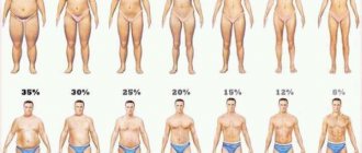
Body fat. Percentage
Fat is connective tissue in the body. First of all, it performs a protective function. Fat is also a reserve source of energy. The fat cells that make up adipose tissue have their own unique structure, which is completely different from the protein cells (that make up muscle tissue).
- For women who do not play sports, the following fat content is usually considered normal: 16-20% and 20-21% (age taken into account).
- For men who do not engage in physical activity, the norm is 8-14% and 10-14%.
In general, body fat percentage is actually a person's weight without all the fat in their body.
Fat or weightlifter?
You can often find a common example in the comparison of fat and muscle: a well-fed person can weigh 100 kg and not look very beautiful, and a bodybuilder who also weighs 100 kg, but has a low percentage of fat, nevertheless looks quite aesthetically pleasing. Same weight, but different shape. In the first case, the person will seem much larger in size than the second, but meanwhile they have the same weight, so what is the mystery?
Having understood the question “Which is heavier: muscles or fat in a person,” everyone can clearly understand what actions they need to take depending on their goal of building a figure. After all, only having certain knowledge in a certain matter can you competently approach solving a problem.
So what should we do?
Holidays should be celebrated, but in moderation. You should start your meal with healthy food - lean meat (turkey or chicken). This will leave less room for fatty, sweet pies and cakes. It is good to alternate feasts with periods of moderate nutrition. It is wrong to go on a complete hunger strike after the holidays, but restricting food intake will be useful. Even with a reduction in calories to a quarter of the daily requirement, appetite hormones will not be released one day, so such a restriction will not stimulate gluttony the next day and will not lead to weight gain. On the contrary, intermittent fasting is an effective, proven way to lose weight.
Source:
https://examine.com/
Body Mass Index (BMI)
One of the most common tools for determining your ideal weight in relation to your height is the body mass index (BMI).
To calculate BMI, divide your body weight (in kilograms) by the square of your height (in meters). For example, let's calculate the body mass index for a person weighing 65.5 kilograms and height 183 centimeters:
BMI = 65.5 / (1.83 x 1.83) = 65.5 / 3.3489 = 19.56. It should be remembered that calculating BMI will not give the correct figures for children and pregnant women.
Based on data from the US National Institutes of Health:
- BMI less than 16 – severe underweight;
- BMI from 16 to 18.5 – underweight;
- BMI from 18.5 to 25 – normal weight;
- BMI from 25 to 30 – pre-obesity;
- BMI from 30 to 35 – first degree obesity;
- BMI from 35 to 40 – second degree obesity;
- BMI over 40 is third degree obesity.
Muscle
Many men are now engaged in creating the torso of Apollo from their bodies. Pumped up biceps and buttocks have become a cult in our time. And it’s not surprising, aesthetics is important for a person in everything, and our body is no exception.
So, we have already found out that fat will not develop into muscle. Then where can you get them from?
It is known that muscles grow from physical activity. But it is not so. The muscles themselves do not grow, but only slightly increase in volume. Without certain nutrition, muscle tissue will not grow larger.
What's the problem with BMI?
Calculating BMI is a simple measurement and is widely used. However, the main factor is the person's height and is not taken into account:
- hip and waist measurements;
- features of the figure and fat distribution;
- muscle mass.
These indicators also have an impact in determining normal weight. For example, athletes have high BMIs despite being in excellent physical shape. This is explained by a large amount of muscle mass, and this does not mean being overweight.
BMI rather gives an approximate understanding of whether a person can be called a healthy weight, rather than defining ideal parameters. BMI measurements are also useful for statistics when researchers calculate trends. This indicator should not become the only one when assessing a person’s normal weight. At a minimum, it is worth considering additional factors.

