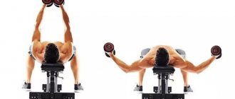
The largest muscles in humans help the body move and lift heavy loads. Muscles and skeleton determine the shape and fit of our body. It is worth noting that they are divided into 3 types: cardiac, skeletal and smooth, and also have differences in their structure and functions. According to different calculation methods, there are from 640 to 850 muscles concentrated in the human body.
Muscles consist of muscle tissue that has the ability to contract under the influence of nerve impulses. They have the ability to convert the energy of chemical reactions occurring in the body into mechanical work. In this article we will look at the largest muscles in the human body.
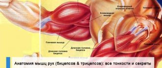
Semimembranosus muscle
The muscle is a posterior thigh muscle. It is responsible for flexing the lower leg at the knee, extending the hip and torso.
In conclusion, I would like to add that, among other things, muscles store energetically valuable material and are a source of heat. If a person has trained muscles, then they can protect the person’s internal organs from various injuries.
The muscle mass of an adult can be up to 40% of his body weight (for leading weightlifters up to 60%), while 86% of it consists of water. Muscle tissue is so important that if a person's muscles suddenly stopped working, he would not even be able to breathe!
0
Share
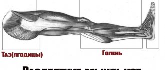
Shin area
Belonging to the muscles of the back of the leg, the gastrocnemius muscle with its superior head is attached to the heel plate by the Achilles tendon. Medially the muscle is attached to the corresponding (medial) femoral condyle, and laterally, respectively, to the lateral one.
The function of the muscle is to provide flexion of the lower leg and foot. In addition, muscle activity is involved in rotational and translational movements of the ankle joint. Particularly active during walking, riding and running. Provides a stable foot position.
The gastrocnemius muscle is formed by the medial and lateral heads. Their direction is symmetrical. However, the medial head is stronger, originating in the popliteal region of the thigh. The first sections of the heads form the lower border of the fossa under the knee joint. They end by coming together in the middle of the lower leg, after which they form the Achilles tendon, which is attached to the posterior tubercle of the heel.
Interesting Facts:
- It is considered the strongest muscle in terms of its ability to stretch. During tension, it can withstand loads of up to 140 kg, depending on the person’s training.
- Shortened in 90% of people. The reasons are underdevelopment of the gluteal muscle and insufficient extension of the foot when walking or running. The danger is moving the center of gravity forward. One of the causes of severe cramps is overstrain of the calf muscle. Injuries often occur due to light but long-term loads.
- A little-known anomaly is the development of the 3rd head of the gastrocnemius muscle. Usually it does not cause discomfort to a person, but it is rare: in about 3% of people. It is more often diagnosed in the Japanese - 5% of occurrence. The third head is attached at the top between the other heads and attached to one of them from the bottom.
Content
- 1 Longissimus thoracis muscle. Lateral tract, sacrospinous system 1.1 Beginning
- 1.2 Attachment
- 1.3 Innervation
- 1.4 Functions
- 1.5 Functional muscle tests
- 2.1 Beginning
- 3.1 Beginning
Heart
A hollow muscular organ that ensures the movement of blood through the vessels. The organ is located in the chest, in the center, and moves slightly to the left. The shape and position of the heart are not the same, and these characteristics are inherited.
Myocardium is cardiac type muscle. Forms the middle layer of the heart, ventricles and atria. The myocardium includes fibers of the atria and ventricles, as well as loose fibrous connective tissue and coronary vessels. It consists of myocytes, which, in turn, are of three types: conductive, contractile and secretory.
The heart muscle is aimed at maintaining homeostasis - the internal environment of the body. The pumping function of the heart is only part of the hydrodynamics (fluid movement) of blood circulation. It is carried out through cyclic contractions and relaxations.
Interesting Facts:
- The most enduring muscle. The myocardium begins to function from birth to death of a person without stopping.
- Factors that lead to a decrease in the tone of the heart muscle are excess calcium and lack of glycogen.
- The myocardium circulates more than 2,500 gallons of blood per day.
- Heart muscle does not have the same ability to regenerate as other muscles.
- Throughout life, the heart makes approximately 2.5 billion contractions.
- Men's heart weighs more than women's. 300-360 and 250-320 respectively.
Reproductive system
The uterus is located in the pelvic cavity of women near the bladder and rectum. Divided into the body and cervix. The normal length for a healthy woman is about 4-6 cm, and the thickness and width are 4-5 cm each. It also depends on the woman’s age and the number of births. The uterus is capable of occupying different positions and is relatively mobile. The muscles are located in the middle layer of the organ walls.
The main function of the uterus is reproductive. This is the ability to fertilize, develop and bear an embryo. The muscles of the uterus ensure that the walls stretch and push out the baby during childbirth.
The uterus consists of spiral-shaped muscle fibers that are directed in different directions. The fundus of the uterus is the upper part of the organ that protrudes above the entrance of the fallopian tubes. The corpus is the triangle-shaped area that tapers toward the cervix.
A peculiarity of the smooth muscles of the uterus is the presence of nexuses - specific gaps between the fibers, which allow the organ to stretch and even contract in this state. Moreover, the contractile function of the uterus largely depends on the concentration of female hormones.
Interesting Facts:
- The uterine muscle is the second in absolute strength after the masticatory muscles.
- In addition to the fact that the uterus stretches greatly during pregnancy, the muscle can contract. During childbirth, the muscle pushes out a child who is several times larger in mass.
The uterus is considered the strongest in the human body, if you take into account the weight of the muscle, despite the fact that the gluteus is the largest.
Author: Svitkevich Julia
Language
The tongue is located in the oral cavity. The muscles make up the main part of the tongue and are divided into 2 groups: skeletal and intrinsic. Both groups include 4 muscles. The muscles of the tongue are symmetrically separated by a longitudinal fibrous septum. All muscles of the tongue are attached to either the hyoid or the mandibular bone.
Among the functions of the tongue are the functions of the papillae located on the mucous membrane of the organ, and the muscles themselves, which make up the bulk of the organ. There are 8 of them in total, each of which performs a specific function.
The skeletal muscles of the tongue include:
- Genioglossus. Provides downward and forward movement of the top of the organ.
- Hypoglossal. Participates in swallowing a bolus of food: it closes the larynx with the epiglottis. Responsible for backward and downward movements.
- Palatoglossus. Provides elevation of the root of the tongue.
- Styllingual. Movement of the tip of the tongue up and back.
All muscles of the second group change the shape of the tongue.
The proper-lingual muscles include:
- Upper longitudinal.
- Lower longitudinal.
- Transverse muscle.
- Vertical muscle.
The tongue is a muscular organ consisting primarily of striated muscles. The organ has the ability to change its shape and position. All muscles of the tongue begin with the occipital myotomas. Therefore, they are innervated only by the 7th pair of cranial nerves.
Interesting Facts:
- The structure of the tongue, called the muscular hydrostat, has been compared to an elephant's trunk.
- Most people believe that the tongue is considered the strongest muscle in the human body, but this is a misconception. The strongest muscle is the chewing muscle.
- Nick Stoeberl had the longest tongue. Its length was 10 cm.
Grade
Find the iliac crest and move back along the ridge to a small bony prominence called the posterior superior iliac spine (PSIS). Place your palm so that your fingers face down and point toward the midline of your body. Now the top of the hand covers the beginning of the muscle, and the bulk of the muscle is located under the palm. This can be confirmed by muscle contraction. The MMN can also be palpated while it is in operation, such as during standing hip extension, platform rise, or medial foot lift.
Treatment
First, you need to understand the root cause of the weakness of the gluteus maximus muscle and try to solve this problem. After this, you can move on to strengthening the muscle using the exercises indicated above (guided by the principle “from simple to complex”).
Development of strength qualities
- Hip extension from a prone position.
- Exercise “Good morning” with weights.
- Stepping onto the platform/bollard.
- Functional tasks (steps, etc.).
Head area
The muscles of mastication belong to the muscles of the head. Together with the facial muscles, they form a complex of the most mobile and sensitive muscles. The structure and tone of the facial muscles influences the shape of the face and determines a person’s tendency to express certain emotions. The activity of facial muscles changes the perception of a person by others.
Anatomically, all masticatory muscles are usually divided into 4 muscles, each of which performs a specific function and is involved in the organization of facial expressions and the primary processing of the food bolus. All 4 muscles are attached to the lower jaw.
In turn, chewables include:
| Name | Location | Function |
| Temporal | It is divided into 2 heads and forms a complex dense tendon resembling a fan. It bends around the zygomatic bone and provides translational movements of the lower jaw | Active during chewing and grinding food. |
| Chewable | Consists of 3 processes: intermediate, superior and inferior. The superficial and lower parts of the masticatory muscle itself begin from the upper and inner surface of the cheekbone, respectively. Both are attached to the lower jaw. The intermediate process originates from the inner surface of the cheekbone and is attached to the outer part of the jaw. | The combined activity of all three processes allows for translational movements of the jaw. Separately, the superficial process provides for the forward movement of the jaw, and the rest for its elevation. Participates in the formation of emotions of dissatisfaction, anger and disgust. |
| Pterygoid lateral | It is formed by two processes and is shaped like a triangle. Located in the infratemporal region. | The activity of two parts of the muscle allows the lower jaw to move forward. In turn, when one of the parts, for example the left, is active, it shifts the lower jaw in the direction opposite to the muscle - to the right. |
| Pterygoid medial | The shape resembles a quadrangle. Located in the lower part of the jaw, under the tongue. Attaches to the pterygoid fossa and tuberosity located on the lower jaw. | Joint contraction ensures the jaw lifts, while relaxation ensures the jaw lowers. Thus, it is active during the grinding of food. Contraction of the right side allows you to move the jaw in the opposite direction. Same with the left side. |
Thus, the chewing muscles are involved in the processes of crushing, grinding and grinding food. It forms the familiar features and proportions of the face, and also helps to express emotions such as anger, fear and strong excitement. Provides both verbal (verbal) and nonverbal (facial) communication.
The largest muscle in the human body is the gluteus maximus, but the muscles of mastication are much stronger and with the muscles of facial expression provide an incredible variety of capabilities.
Interesting Facts:
- The chewing system is capable of creating pressure of up to 100 kg during extreme loads.
- When chopping and grinding ordinary (that is, not liquid, not solid) food, the masticatory muscles create a pressure of 10-15 kg.
- Without exception, all chewing muscles are involved in the act of raising, lowering or displacing the lower jaw, while the upper remains part of the skull and is motionless.
- The peculiarity of the facial muscles is that they are not attached by two heads to the skeleton, but the muscles of mastication are an exception to the rule.