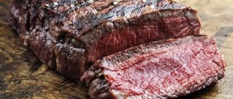
general information
Each muscle, even the smallest one, attached to the bones of the ear, is a separately functioning organ and is controlled by specific nerve endings. Depending on the classification, the number of muscles in the human body may vary - if small muscles are included in larger muscle complexes, then their number is 640.
If we count all muscle tissue (for example, individual cells of smooth muscle tissue) as muscles, then we can count about a billion muscles. Scientists have proposed classifying all types of muscle tissue and classifying cells into individual structures, and those, in turn, into complexes of structures.
Anatomically, muscles are divided not only by location in the body (masticatory, gluteal) and the direction of muscle fibers (directed straight, transversely), but also by the structure of the joints (single-joint muscles, two-joint muscles), as well as by the trajectory of movement (flexion and extension, supinators-pronators ) and taking into account the function they perform (agonists or antagonists).
There are 3 types of muscles in the human body:
- Skeletal muscles. Made up of thousands of small muscle fibers and attached to bones. Only skeletal muscles are subject to voluntary control.
- Smooth muscles. They regulate processes that are unconscious to humans: gastrointestinal peristalsis, inhalation and exhalation, and the functioning of internal organs. Their contractions are involuntary.
- Cardiac muscle. Involuntary, like smooth muscle. Reacts to electrical impulses and provides a heartbeat.
The amount of muscle mass determines the shape of the body. Normally, in a healthy person, muscle weight is up to 45% of body weight. In men, muscle tissue is better developed, formed faster and, accordingly, the percentage of muscles is greater than in women. The toughest people, weightlifters, have more than 60% muscle of their total body weight. This is approximately 40-60 kg of muscle.
The structure of human muscles
The unit of structure of muscle tissue is the muscle fiber. Even a single muscle fiber can contract, indicating that a muscle fiber is not only a single cell, but also a functioning physiological unit capable of performing a specific action.
An individual muscle cell is covered by a sarcolemma.
– a strong elastic membrane provided by the proteins
collagen
and
elastin
. The elasticity of the sarcolemma allows the muscle fiber to stretch, and some people show miracles of flexibility - doing the splits and performing other tricks.
In the sarcolemma, like twigs in a broom, threads of myofibrils
, composed of individual sarcomeres. Thick myosin filaments and thin actin filaments form a multinucleated cell, and the diameter of the muscle fiber is not a strictly fixed value and can vary over a fairly wide range from 10 to 100 microns. Actin, which is part of the myocyte, is an integral part of the cytoskeleton structure and has the ability to contract. Actin consists of 375 amino acid residues, which makes up about 15% of the myocyte. The remaining 65% of muscle protein is myosin. Two polypeptide chains of 2000 amino acids form the myosin molecule. When actin and myosin interact, a protein complex is formed - actomyosin.
Description of human muscles
difficult, and for a visual representation you can refer to the textbook “Grade 8 Biology” edited by V.I. Sivoglazov, where on page 117 the illustration shows what a myocyte looks like under a microscope.
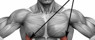
The most powerful
The gluteus maximus muscle (or GLM) is rightfully the largest in the human body.
In the lower segment of the body there are 3 gluteal muscles: small, medium and large. The latter is shaped like a diamond and is attached to the ilium, sacrum, and coccyx. It stretches from the side of the spine along the pelvis and attaches to the femur on the other side.
The gluteus is the largest muscle in the human body
The main function is hip extension during high loads. The BNM is responsible for the relative mobility of the femoral fascia to which it is attached. Fascia is a mobile extension of connective tissue from the femur.
The largest muscle in the human body is responsible for squatting, maintaining posture, walking, stair stepping, bending to the side and running.
The gluteus maximus muscle is a fleshy, dense mass of skeletal muscle. In athletes, the muscle reaches 2-3 cm in thickness. Consists of bundles of parallel-lying rough muscle fibers.
Interesting Facts:
- The gluteus maximus muscle is the most massive and largest muscle in the human body.
- The size of the muscle is characteristic of erect walkers in general, since it supports the torso in an upright position.
- It has great aesthetic significance as it lies above the smaller gluteal muscles and provides the shape of the buttocks.
What is the function of the gluteus maximus muscle?
The human body is constantly in motion. He has to take on a variety of body loads, and sometimes we don’t even suspect how much weight falls on him.
Thanks to the gluteal muscles, a person can successfully perform work such as climbing stairs, carrying large objects, and rearranging furniture. During this type of work the following muscles are involved:
- gluteus maximus;
- femoral;
- and also responsible for straightening the spine.
Athletes need the gluteus maximus muscle to perform a huge range of exercises. A young athlete is often faced with the need to develop their body in a comprehensive manner. The muscle in question is needed to perform jerking movements performed when running, jumping, throwing a discus (hammer) and in other disciplines. It takes on heavy loads during swimming, when the swimmer chooses freestyle. It has to withstand an even greater load when climbing uphill, and the muscles of the body experience the most serious loads during wrestling, when wrestlers have to put in the strength of their legs. Baseball and badminton players use it the moment they start swinging their bat or racket.
American football players also often turn their feet to throw the ball. Many boxers try to use all the strength of their gluteus maximus muscle to deliver the most powerful punch possible.
Thus, no matter what sport you take, it is of paramount importance. Moreover, this applies not only to dynamic sports, but also to power sports. For example, powerlifting and weightlifting athletes perform compound exercises such as barbell squats and deadlifts. They often have to switch to jerks, using all the reserves of their body. This muscle absorbs the load, absorbs impulses, and performs isometric work. Of course, other muscles also help her, for example, the thigh muscles. That is why their development and training are extremely important to achieve successful results.
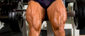
Femoral area
The sartorius muscle is attached at one end to the ilium, bending around the surface of the thigh, and ends at the anteromedial region of the leg. Forms a dense tendon. The superior head of the sartorius muscle is the border of the femoral triangle.
This muscle provides inward and outward movement of the lower leg and thigh joints. Actively participates in raising the leg, straightening the hip, and helps maintain body posture. Helps maintain balance when performing squats.
It belongs to the muscles of the anterior thigh, along with the quadriceps femoris and the articular muscle of the knee. Innervated by the femoral nerve. The blood supply is carried out by the muscular branches of the femoral artery.
The largest muscle in the human body is easily palpated and even visually observed during abduction of the legs to the side or extension of the limb.
Interesting Facts:
- The longest muscle in the human body. The average adult reaches 43-44 cm.
- The functions of the sartorius muscle allow a person to sit cross-legged on a hard surface.
- The sartorius muscle of the left leg is more active if, simultaneously with the movements, manipulations are carried out with the opposite upper limb and vice versa.
- It is called “sartorial” because it bends the hip in a similar way to how tailors do it during the work process.
Shin area
Belonging to the muscles of the back of the leg, the gastrocnemius muscle with its superior head is attached to the heel plate by the Achilles tendon. Medially the muscle is attached to the corresponding (medial) femoral condyle, and laterally, respectively, to the lateral one.
The function of the muscle is to provide flexion of the lower leg and foot. In addition, muscle activity is involved in rotational and translational movements of the ankle joint. Particularly active during walking, riding and running. Provides a stable foot position.
The gastrocnemius muscle is formed by the medial and lateral heads. Their direction is symmetrical. However, the medial head is stronger, originating in the popliteal region of the thigh. The first sections of the heads form the lower border of the fossa under the knee joint. They end by coming together in the middle of the lower leg, after which they form the Achilles tendon, which is attached to the posterior tubercle of the heel.
Interesting Facts:
- It is considered the strongest muscle in terms of its ability to stretch. During tension, it can withstand loads of up to 140 kg, depending on the person’s training.
- Shortened in 90% of people. The reasons are underdevelopment of the gluteal muscle and insufficient extension of the foot when walking or running. The danger is moving the center of gravity forward. One of the causes of severe cramps is overstrain of the calf muscle. Injuries often occur due to light but long-term loads.
- A little-known anomaly is the development of the 3rd head of the gastrocnemius muscle. Usually it does not cause discomfort to a person, but it is rare: in about 3% of people. It is more often diagnosed in the Japanese - 5% of occurrence. The third head is attached at the top between the other heads and attached to one of them from the bottom.
Head area
The muscles of mastication belong to the muscles of the head. Together with the facial muscles, they form a complex of the most mobile and sensitive muscles. The structure and tone of the facial muscles influences the shape of the face and determines a person’s tendency to express certain emotions. The activity of facial muscles changes the perception of a person by others.
Anatomically, all masticatory muscles are usually divided into 4 muscles, each of which performs a specific function and is involved in the organization of facial expressions and the primary processing of the food bolus. All 4 muscles are attached to the lower jaw.
In turn, chewables include:
| Name | Location | Function |
| Temporal | It is divided into 2 heads and forms a complex dense tendon resembling a fan. It bends around the zygomatic bone and provides translational movements of the lower jaw | Active during chewing and grinding food. |
| Chewable | Consists of 3 processes: intermediate, superior and inferior. The superficial and lower parts of the masticatory muscle itself begin from the upper and inner surface of the cheekbone, respectively. Both are attached to the lower jaw. The intermediate process originates from the inner surface of the cheekbone and is attached to the outer part of the jaw. | The combined activity of all three processes allows for translational movements of the jaw. Separately, the superficial process provides for the forward movement of the jaw, and the rest for its elevation. Participates in the formation of emotions of dissatisfaction, anger and disgust. |
| Pterygoid lateral | It is formed by two processes and is shaped like a triangle. Located in the infratemporal region. | The activity of two parts of the muscle allows the lower jaw to move forward. In turn, when one of the parts, for example the left, is active, it shifts the lower jaw in the direction opposite to the muscle - to the right. |
| Pterygoid medial | The shape resembles a quadrangle. Located in the lower part of the jaw, under the tongue. Attaches to the pterygoid fossa and tuberosity located on the lower jaw. | Joint contraction ensures the jaw lifts, while relaxation ensures the jaw lowers. Thus, it is active during the grinding of food. Contraction of the right side allows you to move the jaw in the opposite direction. Same with the left side. |
Thus, the chewing muscles are involved in the processes of crushing, grinding and grinding food. It forms the familiar features and proportions of the face, and also helps to express emotions such as anger, fear and strong excitement. Provides both verbal (verbal) and nonverbal (facial) communication.
The largest muscle in the human body is the gluteus maximus, but the muscles of mastication are much stronger and with the muscles of facial expression provide an incredible variety of capabilities.
Interesting Facts:
- The chewing system is capable of creating pressure of up to 100 kg during extreme loads.
- When chopping and grinding ordinary (that is, not liquid, not solid) food, the masticatory muscles create a pressure of 10-15 kg.
- Without exception, all chewing muscles are involved in the act of raising, lowering or displacing the lower jaw, while the upper remains part of the skull and is motionless.
- The peculiarity of the facial muscles is that they are not attached by two heads to the skeleton, but the muscles of mastication are an exception to the rule.
Language
The tongue is located in the oral cavity. The muscles make up the main part of the tongue and are divided into 2 groups: skeletal and intrinsic. Both groups include 4 muscles. The muscles of the tongue are symmetrically separated by a longitudinal fibrous septum. All muscles of the tongue are attached to either the hyoid or the mandibular bone.
Among the functions of the tongue are the functions of the papillae located on the mucous membrane of the organ, and the muscles themselves, which make up the bulk of the organ. There are 8 of them in total, each of which performs a specific function.
The skeletal muscles of the tongue include:
- Genioglossus. Provides downward and forward movement of the top of the organ.
- Hypoglossal. Participates in swallowing a bolus of food: it closes the larynx with the epiglottis. Responsible for backward and downward movements.
- Palatoglossus. Provides elevation of the root of the tongue.
- Styllingual. Movement of the tip of the tongue up and back.
All muscles of the second group change the shape of the tongue.
The proper-lingual muscles include:
- Upper longitudinal.
- Lower longitudinal.
- Transverse muscle.
- Vertical muscle.
The tongue is a muscular organ consisting primarily of striated muscles. The organ has the ability to change its shape and position. All muscles of the tongue begin with the occipital myotomas. Therefore, they are innervated only by the 7th pair of cranial nerves.
Interesting Facts:
- The structure of the tongue, called the muscular hydrostat, has been compared to an elephant's trunk.
- Most people believe that the tongue is considered the strongest muscle in the human body, but this is a misconception. The strongest muscle is the chewing muscle.
- Nick Stoeberl had the longest tongue. Its length was 10 cm.
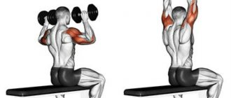
Antagonist muscles and synergist muscles
In modern bodybuilding, most training programs are built taking into account the conditional division of muscles into antagonists and synergists. Antagonists are muscle groups that create the opposite effect in relation to each other, that is, in other words, these are the flexor and extensor muscles of the joints. While performing an exercise on a specific muscle, the opposite antagonist is in the stage of rest or light static tension. Thus, training can be built on the principle of working out muscles in pairs, taking into account their size and recovery ability. The main paired muscle groups of antagonists: Biceps - triceps Quadriceps - hamstrings Pectoral muscles - latissimus dorsi Synergists are muscle groups that work unidirectionally, i.e. perform the same contractile function in various exercises. The principle of training synergist muscles is to work large muscle groups in combination with small or minor ones. This applies to multi-joint exercises that involve both, as well as individual movements targeting secondary muscles. The main paired muscle groups of synergists: Triceps - pectoral muscles Latissimus dorsi - biceps Leg muscles - buttocks Shoulders (deltoid bundles) are considered to be synergists, since their development has several directions - mainly in bench presses, as well as in all kinds of rows and extensions at different angles. WHAT IS IT MORE EFFECTIVE TO TRAIN? There are still a lot of opinions and disagreements about what muscles to train and how to create a schedule. Despite the abundance of different options for split programs, it is impossible to determine exactly which workout will be effective for a particular person. But if we do not take into account the structural features of the body of all bodybuilders, and take, say, two people of similar physique of the same height and weight, we can conduct an experiment that will give a more or less accurate answer to our question. We will not consider what type of work will be performed in class. Both athletes will work for the entire month according to the same training scheme (antagonists), spending the same amount of time on work, as well as on rest between approaches and exercises. It would be fair to add to all this the requirement of observing the correct movement technique. The result can be confusing in the first weeks. One athlete will progress confidently, while the other will perhaps get some result, but it will be insignificant. Most likely, the second athlete will remain in the same place, and in the future will drive himself into a state of overtraining. Thus, it is obvious that the recovery ability of athletes is different, and it’s not even a matter of the amount of rest or sleep. It’s just that athletes have individual hormonal backgrounds, different rates of recovery of adenosine triphosphoric acid, glycogen and the muscle fibers themselves. Based on this, the second athlete can change direction from antagonist training to synergist training and further progress as quickly as the first athlete. HOW TO COMPETENTLY CREATE A PROGRAM Knowing your own predisposition to the work of antagonists and synergists, it is important to understand the simple rule for arranging the sequence of exercises, which will later help you create your own program. In any classical scheme, training begins with performing exercises for large muscle groups - the latissimus dorsi, pectoral muscles, quadriceps and hamstrings. The legs have the largest muscle group, so it is recommended to dedicate a separate training day for them. When training a large synergist muscle, remember that at the same time, the small (minor) muscle is also “working”, which quickly becomes clogged. Therefore, it is advisable to work with the small one at the end of the workout, and not vice versa. Otherwise, for example, a tired biceps will not allow you to fully and efficiently work the latissimus dorsi muscle in pull-ups, and the triceps will not allow you to work out the pectoral muscle in presses. In the case of antagonists, it makes sense to split the weekly split into training the back, chest and legs at the beginning of the week, and finish with work on the biceps and triceps at the end. You can break up your weekly program even further by dedicating one day to each muscle group. This version of the split will allow you to better work out a specific muscle, focusing on its individual bundles. To summarize the above, antagonists and synergists are a conditional division of muscles into groups in order to create the correct (read, suitable for you) program. To understand what and how best to train, there is no definite answer - to understand the principles of successful growth and rapid progression may take more than one week, or even months. It is important to understand that you first need to determine your predisposition to a particular type of training based on the body’s recovery ability. In addition, for good progress, it is absolutely necessary to fully take into account all factors - rest (including keeping stress to a minimum), nutrition and, in fact, the training process itself.
Heart
A hollow muscular organ that ensures the movement of blood through the vessels. The organ is located in the chest, in the center, and moves slightly to the left. The shape and position of the heart are not the same, and these characteristics are inherited.
Myocardium is cardiac type muscle. Forms the middle layer of the heart, ventricles and atria. The myocardium includes fibers of the atria and ventricles, as well as loose fibrous connective tissue and coronary vessels. It consists of myocytes, which, in turn, are of three types: conductive, contractile and secretory.
The heart muscle is aimed at maintaining homeostasis - the internal environment of the body. The pumping function of the heart is only part of the hydrodynamics (fluid movement) of blood circulation. It is carried out through cyclic contractions and relaxations.
Interesting Facts:
- The most enduring muscle. The myocardium begins to function from birth to death of a person without stopping.
- Factors that lead to a decrease in the tone of the heart muscle are excess calcium and lack of glycogen.
- The myocardium circulates more than 2,500 gallons of blood per day.
- Heart muscle does not have the same ability to regenerate as other muscles.
- Throughout life, the heart makes approximately 2.5 billion contractions.
- Men's heart weighs more than women's. 300-360 and 250-320 respectively.
Reproductive system
The uterus is located in the pelvic cavity of women near the bladder and rectum. Divided into the body and cervix. The normal length for a healthy woman is about 4-6 cm, and the thickness and width are 4-5 cm each. It also depends on the woman’s age and the number of births. The uterus is capable of occupying different positions and is relatively mobile. The muscles are located in the middle layer of the organ walls.
The main function of the uterus is reproductive. This is the ability to fertilize, develop and bear an embryo. The muscles of the uterus ensure that the walls stretch and push out the baby during childbirth.
The uterus consists of spiral-shaped muscle fibers that are directed in different directions. The fundus of the uterus is the upper part of the organ that protrudes above the entrance of the fallopian tubes. The corpus is the triangle-shaped area that tapers toward the cervix.
A peculiarity of the smooth muscles of the uterus is the presence of nexuses - specific gaps between the fibers, which allow the organ to stretch and even contract in this state. Moreover, the contractile function of the uterus largely depends on the concentration of female hormones.
Interesting Facts:
- The uterine muscle is the second in absolute strength after the masticatory muscles.
- In addition to the fact that the uterus stretches greatly during pregnancy, the muscle can contract. During childbirth, the muscle pushes out a child who is several times larger in mass.
The uterus is considered the strongest in the human body, if you take into account the weight of the muscle, despite the fact that the gluteus is the largest.
Author: Svitkevich Julia
What actions should a person take when the musculus sartorius is stretched?
When direct compression of the fibers or severe stretching occurs, the innervation of the musculus sartiorius is disrupted.
In some cases, neuropathy occurs due to diabetes. It is imperative to contact a neurologist, who should conduct the most thorough examination and electrodiagnostics. Additionally, the patient must be prepared for tomography and MRI. Despite its apparent simplicity, even many large muscles do not require such complex treatment as the longest one. Typically, the patient is prescribed medication, relaxation procedures, and fiber stretching. All work will require concentration on correcting only those muscles that are associated with the damaged area.