| Internal oblique muscle | |
| Internal oblique abdominal muscle. | |
| Muscles of the trunk. | |
| details | |
| Origin | Inguinal ligament, Iliac crest and psoas fascia. |
| Insert | linea alba, thoracic line pubis (through the fused tendon) and ribs 10-12. |
| Artery | Subcostal arteries. |
| Nerve | Thoracoabdominal nn. (T7-T11), Subcostal n. (T12), Iliohypogastric n. (L1) and Ilioinguinal n. (L1) |
| Actions | Bilateral: compresses the abdomen, unilateral: ipsilateral rotation of the trunk. |
| Identifiers | |
| Latin | Musculus obliquus internus abdominis |
| TA98 | A04.5.01.017 |
| TA2 | 2373 |
| F.M.A. | 13891 |
| Anatomical conditions of the muscle [edit in Wikidata] | |
The internal oblique muscle
, also
the internal oblique muscle
or
oblique internal muscle
, is an abdominal muscle in the abdominal wall that lies below the external oblique muscle and just above the transverse abdominis muscle.
Structure
Its fibers run perpendicular to the external oblique muscle, starting from the thoracolumbar fascia of the lower back, the anterior 2/3 of the iliac crest (upper part of the femur) and the lateral half of the inguinal ligament. Muscle fibers pass from these points superiomedially (up and towards the midline) to the muscle attachments on the lower borders of the 10th to 12th ribs and to the muscles. linea alba.
In men, the cream muscle is also attached to the internal oblique.
Nervous nutrition
The internal oblique muscle is supplied by the inferior intercostal nerves, as well as the iliohyoid nerve and the ilioinguinal nerve.
Muscles that form the anterior abdominal wall
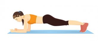
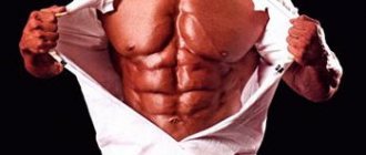
1. Rectus abdominis (musculus rectus abdominis). Things didn’t work out with high-quality cadaveric material at my university, so I saw this muscle for the first time during an operation (it was my third year, I think). It looks like a fairly wide strap that stretches strictly vertically across the entire stomach.
More precisely, two wide straps, because the muscle is steamy.
The rectus abdominis muscle is divided by tendon bridges (intersectiones tendinae), which are located across the fibers of the muscle. You can see the white cross stripes in both illustrations. It is these jumpers that form the “cubes” that are clearly visible in athletic people.
The rectus abdominis muscle is enclosed in the vagina (vagina musculi recti abdominis), which consists of aponeuroses of the muscles of the lateral abdominal wall.
Origin: pubic bone, the space between the pubic syphysis and the pubic tubercle.
Insertion: anterior surface of the xiphoid process of the sternum; outer surface of the cartilage of 5-7 (inclusive) ribs.
Function: bends the spine, with the chest fixed (for example, hanging on a horizontal bar), raises the pelvis.
2. Pyramidal muscle (musculus pyramidalis)
This is a rather small muscle, it is not always possible to see it. To see it, you need to look for a small, triangular-shaped muscle in the pubic area. In the figure above, in which we noted the rectus abdominis muscle, we can also find the pyramidal muscle:
Origin: pubic bone, anterior to the beginning of the rectus abdominis muscle;
Attachment: lower part of the white line of the abdomen;
Function: tightens the linea alba.
Feature: the muscle is vestigial. According to Sapin's textbook, the pyramidalis muscle may be completely absent.
Function
The internal oblique element performs two main functions. First, as an accessory muscle of respiration, it acts as an antagonist to the diaphragm, helping to reduce the volume of the chest cavity during exhalation. When the diaphragm contracts, it pulls down the lower wall of the chest, increasing the volume of the lungs, which then fill with air. Conversely, when the internal obliques contract, they compress the abdominal organs, pushing them upward toward the diaphragm, which returns back into the chest cavity, reducing the volume of the air-filled lungs, causing exhalation.
Secondly, its contraction causes ipsilateral rotation and lateral bending. It acts with the external oblique muscle of the opposite side to achieve this torsional movement of the trunk. For example, the right internal oblique and left external oblique muscles contract when the torso flexes and rotates to bring the left shoulder closer to the right hip. For this reason, the internal inclined parts are called "one-sided rotators."
Additional images
- Diagram of a cross-section of the posterior abdominal wall showing the location of the psoas fascia.
- Diagram of the vagina of the rectum.
- Cross-sectional diagram of the anterior abdominal wall below the semicircular line.
- The shell of the femur is opened to show its three compartments.
- Lumbar triangle
- Internal oblique abdominal muscle. Anterior abdominal wall. Deep dissection. Front view.
Rice. 115. Muscles and fascia of the trunk. Front view. 1-fascia of the chest (superficial layer); 2-deltoid-pectoral groove; 3-deltoid fascia; 4-fascia of Gshech; 5-fascia of the abdomen; 6-umbilical ring; 7-superior anterior iliac spine; 8-sperm cord; 9-inguinal ligament; 10-blue line of the abdomen; 11-aponeurosis of the external oblique abdominal muscle; 12-external oblique abdominal muscle; 13-serratus anterior muscle; 14 latissimus dorsi muscle; 15-biceps brachii; 16-major muscle; 17 deltoid muscle; 18-clavicle; 19-fudino-cleidomastoid muscle; 20-subcutaneous muscle of the neck.
Fig. 115. Muscles and fascia of the trunk. Front view. 1-fascia pectoralis (lamina superficialis); 2-sulcus deltoideopectoralis; 3-fascia deltoidea; 4-fascia brachii; 5-fascia propria abdominis (lamina superficialis); 6-anulus umbilicalis; 7-spina iliaca anterior superior; 8-funiculus spermaticus; 9-lig.inguinale; 10-linea alba (abdominis); II-aponeurosis m. obliqui cxterni abdominis; 12 am. obliquus externus abdominis; 13.00 serratus anterior; 14-m. latissimus dorsi; 15-m.biceps brachii; 16-m. pectoralis major; 17-m. deltoideus; 18-clavic-ula; 19-m. sternocleidomastoideus; 20-platysma.
Fig. 115. Muscles and fasciesof (rank. Anterior aspect. 1-pectoral fascia (superficial layer); 2-deltoid pectoral groove; 3-del-toid fascia; 4-brachial fascia; 5-proprial abdominal fascia (superficial layer); 6- umbilical ring; 7-anterior superior iliac spine; 8-spermatic cord; 9-inquinal ligament; 10-linea alba (of abdomen); 1 l-aponeurosis of external oblique muscle (of abdomen); 12-external oblique muscle; 13- serratus anterior; 14-latissimus dorsi; 15-biceps brachii; 16-pectoral major; 17-deltoid; 18-clavicule; 19-sternoclcidomastoid; 20-platysma.
Rice. 116. Muscles of the trunk. Right view.
1-pectoralis major muscle; 2-serratus anterior muscle; 3-external oblique abdominal muscle: 4-aioneurosis of the external oblique abdominal muscle; 5-muscle that strains the fascia lata (hips); 6-gluteus maximus, 7-latissimus dorsi; 8-teres major muscle; 9-teres minor muscle; 10 infraspinatus muscle; 11 deltoid muscle; 12-trapezius muscle; 13-sternocleidomastoid muscle.
Fig. 116. Muscles of the trunk. Right view.
1-m. pectoralis major; 2-vol. serratus anterior; 3-m. obliquus extemus abdominis; 4-aponeurosis m. obliqui extern! abdominis; 5-m.tensor fasciae latae; 6-m. gluteus maximus; 7-m.latissimus dorsi; 8-m. teres major; 9 am. teres minor; 10 am. infraspinatus; 11 am. deltoideus; 12 am. trapezius; 13-rn. sternocleidomastoideus.
Fig. 116. Muscles of trunk. Right aspect.
1-pectoral major; 2-serratus anterior; 3-external oblique (of abdomen); 4-aponeurosis of external oblique (of abdomen); 5-tensor fasciae latae muscle; 6-gluleus maximus; 7-latissimus dorsi; 8-teres major; 9-teres minor; 10-infraspinatus; 11-deltoid; 12-trape/ius; 13-sternocleido-mastoid.
Rice. 117. Muscles of the trunk (food and abdomen). Front view. 1-deep plate of the pectoral fascia; 2-deltoid muscle (pulled to the side); 3-pectoralis major muscle (partially removed); 4-serratus anterior muscle; 5-internal intercostal muscles; 6-rectus abdominis muscle; 7-tendon jumpers; 8-transverse abdominal muscle; 9-internal oblique muscle (cut off and turned down); 10-pyramidal muscle; 11-inguinal ligament; 12-aponeurosis of the internal oblique abdominal muscle; 13-internal oblique abdominal muscle; 14 white line of the abdomen; 15-biceps brachii; 16-minor muscle; 17-major muscle.
Fig. 117. Muscles of the trunk (food and abdomen). Front view. I-lamina profunda fasciae pectoralis; 2-m. deltoideus (pulled to the side); 3-m. pectoralis major (partially removed); 4-m. serratus anterior; 5-mm. intercostales interni; 6-m. rectus abdominis; 7-inter-sections tendineae; 8-m. transversus abdominis; 9 am. obliquus internus abdominis (cut off and turned down); 10 am. pyramidalis; 11-lig. inguinal; 12-aponeurosis m. obliqui interni abdominis; 13.00 obliquus internus abdominis; 14-linea alba (abdominis); 15-m.biceps brachii; 16-m. pectoralis minor; 17-m. pectoralis minor.
Fig. 117. Muscles oftrank (of chest and abdomen). Anterior aspect. 1-pectoral fascia (deep layer); 2-deltoid muscle (displaced); 3-pec-toralis major (partly cut away); 4-serratus anterior; 5-internal intercostal; 6-rectus abdominis; 7-tendinous intersections; 8-lransversus abdominis; 9-internal oblique (cut away and displaced); 10-pyrami-dalis; 11-inquinal ligament; 12-aponeurosis of external oblique (of abdomen); 13-internal oblique (of abdomen); 14-linea alba; 15-biceps brachii; 16-pectoralis minor; 17-pectoralis major.
Rice. 118. Deep muscles of the chest and abdomen. I-internal intercostal muscles; 2-external intercostal muscles; 3-rectus abdominis muscle (partially removed); 4-white line of the abdomen; 5-vagina of the rectus abdominis muscle (posterior plate); 6-lunar line; 7-transverse abdominal muscle; 8th anterior plate of the rectus abdominis sheath; 9-inguinal ligament; 10-sperm cord; 11-pyramidalis muscle (cut off and partially removed); 12-porteur muscle; 13-tensor muscle of the fascia lata (thigh); 14-transversalis fascia; 15-blowing line; 16-serratus anterior muscle; 17 biceps brachii; 18 latissimus dorsi; 19-minor muscle; 20 coracobrachialis muscle; 21 subclavian muscle; 22-trapezius muscle; 23-sternocleidomastoid muscle.
Fig. 118. Deep muscles of the chest and abdomen. 1-mm. intercostalis intemi; 2-mm. intercostalis extern!; 3-m. rectusabdo-minis (partially removed); 4-linea alba (abdominis); 5-vagina m. recti abdominis (lamina posterior); 6-linea arcuata; 7-m. transversus abdominis; 8-vaginae m. recti abdominis (lamina anterior); 9-lig.inguinale; 10-funiculus spermaticus;ll-m. pyramidalis;12-m.sartorius;13-m.tensor tas-ciae latae; 14-fascia transversalis; 15-vagina m.recti abdominis (lamina posterior); 16-m. serratus anterior; 17-m. biceps brachii; 18-m. latissimus dorei; 19-m. pectoralis minor; 20-m. coracobrachialis; 21-m.subclavius; 22-m. trapezius; 23-m. sternocleidomastoideus.
Fig. 118. Deep muscles of thorax and abdomen. 1-internal intercostal muscles; 2-external intercostal muscles; 3-rectus abdominis (partly cut away); 4-linea alba; 5-rectus sheath (posterior layer); 6-arcuate line; 7-transversus abdominis muscle; 8-rectus sheath (anterior layer); 9-inquinal ligament; 10-spermatic cord; 11-pyrami-dalis muscle (cut away); 12-sartorius; 13-tensor fascia latae muscle; 14-fascia transversalis; 15-rectus sheath (posterior layer); 16-serratus anterior muscle; 17-biceps brachii muscle; 18-latissimus dorsi muscle; 19-pectoralis minor muscle; 20-coraco-brachial muscle; 21-subclavius muscle; 22-trapezius muscle; 23-sternocleidomastoid muscle.
Rice. 119. Muscles and fascia of the anterior chest and abdominal walls. Back view.
1 - internal intercostal muscles; 2-transverse chest muscle; 3-transverse fascia; 4-deep inguinal ring; 5-iliopsoas muscle; 6-sperm cord (cut off); 7-external iliac artery; 8-external iliac vein; 9-co-vascular lacuna; 10-iliopectineal arch; 11-deep inguinal ring; 12-muscle lacuna; 13-rectus abdominis muscle; 14-arcuate line; 15 semilunar line; 16-transverse abdominal muscle; 17-diaphragm (costal part).
Fig. 119. Muscles and fascia of the anterior chest and abdominal walls. Back view.
1-mm.intercostales interni; 2-m. transversus thoracis; 3-fascia transversalis; 4-anulus inguinalis profundus; 5-m. iliopsoas; 6-funiculus spermaticus (cut off); 7-a. iliaca externa; 8-v.iliac externa; 9-lacu-na vasorum; 10-arcus iliopectineus; 11-anulus inguinalis profundus; 12-lacuna musculorum; 13.00 rectus abdominis; 14-lineaarcuata; 15-linea semilunaris; 16-m. transversus abdominis; 17-diaphragma (pars costal is).
Fig. 119. Muscles and fascial of anterior thoracis and abdominal walls.
Posterior aspect.
1-internal intercostal muscles; 2-transversus thoracis; 3-fascia transversalis; 4-deep inguinal ring; 5-iliopsoas; 6-spermatic cord (cut away); 7-external iliacal artery; 8-external iliacal vein; 9-vascular space; 10-iliopectineal arch; 11-superficial inguinal ring; 12-muscular space; 13-rectus abdominis (of abdomen); 14-arcuate line; 15-linea semilunaris; 16-transversus abdominis; 17-diaphragm (costal part).
Rice. 120. Diaphragm. View from above. 1st lumbar part of the diaphragm; 2-lumbocostal triangle; 3-costal part of the diaphragm; 4-aorta (aortic opening); 5-esophagus (esophageal opening); 6-sternocostal triangle; 7-sternal part of the diaphragm; 8-tendon center of the diaphragm; 9-inferior vena cava (opening of the inferior vena cava).
Fig. 120. Diaphragma. View from above.
1-pars lumbalis diaphragmae; 2-trigonum lumbocostale; 3-pars costal-is diaphragmae; 4-aorta (hialus aorticus); 5-esophagus (hiatus esophgeus); 6-trigonum sternocostale;7-pars stemalis diaphragmae; 8-centrum tendineum diaphragma; 9-v. cava inferior (foramen vanae cavae).
Fig. 120. Diaphragm.
1-lumbar part; 2-sternocostal triangle; 3-costal part; 4-aorta (aortic hialus); 5-oesophagus (oesophageal hiatus); 6-central tendon; 7-interi-or vena cava (caval opening).
Rice. 121. Diaphragm and muscles of the posterior abdominal wall.
Front view. l-fudinary part of the diaphragm; 2-sternocostal triangle; 3-tendon center of the diaphragm; 4-costal part of the diaphragm; 5-hole of the inferior vena cava; 6-esophageal opening; 7-from the opening of the aorta; 8-left leg of the lumbar part of the diaphragm; 9-lumbocostal triangle; 10 quadratus lumborum muscle; 11-psoas minor muscle; 12-major lumbar muscle; 13-iliac muscle; 14-iliac fascia; 15-iodine ring (femoral canal); 16-external obturator muscle; 17-iliopsoas muscle; 18 psoas major muscle (cut off); 19-ileum mouse; 20-intra-abdominal fascia; 21-intertransverse muscles; 22-medial crus of the diaphragm (left side); 23-medial crus of the diaphragm (right side); 24-lateral arcuate ligament (lateral lumbocostal arch); 25-medial arcuate ligament (medial lumbocostal arch); 26-right leg of the lumbar part of the diaphragm; 27-median arcuate ligament; 28-lumbar part of the diaphragm.
Fig. 121. Diaphragm and muscles of the posterior abdominal wall.
Front view. 1-pars sternalis diaphragmae; 2-trigonum sternocostale; 3-centrum tentiineum diapliragmae; 4-pars costal is diaphragmae; 5-foramen venae cavae (inferioris); 6-hiatus esophageas; 7-hiatus aorticus; 8-crus sinister (partis lumbalis diaphragmae); 9-trigonum lumbocostale; 10 am. quadratic lumborum; 11 am. psoas minor; 12 am. psoas major; 13.00 iliacus; 14-fas-cia iliaca; 15-hiatus saphenus; 16-m. obturatorius externus; 17-m. iliop-soas; 18-m. psoas major (cut off); 19-m. iliacus; 20-fascia endoabdom-inalis; 21-mm. intertransversarii; 22-crus mediale diaphragmae (pars sinistra); 23-crus mediale diaphragmae (pars dextra); 24-lig. arcuatum lal-erale; 25-lig. arcuatum mediale; 26-crus dextrum partis lumbalis diaphragmae; 27-lig. arcuatum medianum; 28-pars lumbalis diaphragmae.
Fig. 121. Diaphragm and muscles of posterior abdominal wall.
Anterior aspect.
1-sternal pan (of diaphragm); 2-sternocostal triangle; 3-central tendon; 4-costal part (of diaphragm); 5-caval opening; 6-oesophageal hiatus; 7-aor tic hiatus; 8-left cms oflumbar part of diaphragm; 9-lumbocostal triangle; I0-quadratuslumborurn; 11-psoas minor; 12-psoas major; 13-iliac muscle; 14-fascia iliaca; 15-subcutancus opening (of femoral canal); 16-obtu-rator extcrnus; 17-iliopsoas; 18-psoas major muscle (cut away); 19-iliac muscle; 20-endoabdominal fascia; 21 -intertransversarii musclca; 22-medi-al crus of diaphragm (left part); 23-medial CRIS of diaphragm (right part); 24-lateral arcuate ligament (lateral lumbocostal arch); 25-medial arcuate ligament (medial lumbocostal arch); 26-right CRIS of lumbar part of diaphragm; 27-middle arcuate ligament; 28-lumbar part of diaphragm.